4231
Regional Brain Volume Changes in Alcohol-dependent Individuals during Short-term and Long-term Abstinence1Department of Radiology and Biomedical Imaging, University of California San Francisco, San Francisco, CA, United States, 2Center for Imaging of Neurodegenerative Diseases, San Francisco VA Medical Center, San Francisco, CA, United States, 3Department of Psychiatry and Behavioral Sciences, Stanford University, Stanford, CA, United States, 4Mental Illness Research and Education Clinical Centers and Sierra-Pacific War Related Illness and Injury Study Center, VA Palo Alto Health Care System, CA, United States
Synopsis
The general goal of this study is to determine the volume changes of cortical and subcortical brain regions in smoking and currently non-smoking alcohol-dependent individuals during short-term and long-term abstinence from alcohol, compared with non-/light-drinking controls. Preliminary results from paired t-tests within each group show that the anterior cingulate cortex, dorsolateral prefrontal cortex, orbitofrontal cortex, insula, and hippocampus have different recovery patterns, which suggest potentially different neuropathological changes and/or injuries. More statistical analyses will test for cross-sectional and longitudinal differences between the groups for a better interpretation of the regional volume changes and their cognitive and behavioral correlates.
INTRODUCTION:
The development and reinforcement of alcohol use disorders involves deficits in complex functional neuro-circuits that are modulated by brain regions affiliated with reward, motivation, stress, memory, and executive control. Critical brain regions include the anterior cingulate cortex (ACC), dorsolateral prefrontal cortex (DLPFC), orbitofrontal cortex (OFC), insula, and hippocampus.1,2 Cross-sectional and longitudinal MR structural studies showed that compared to non-/light-drinking controls (CON), alcohol-dependent individuals (ALC) have a smaller frontal gray matter in early abstinence,3 and that lobar volume changes are greater over short-term than long-term abstinence.4 In ALC, cigarette smoking is also associated with significant morphological abnormalities.5,6 However, the regional volume reductions and their changes within the major lobes of ALC remain unclear. The specific goal of this preliminary analysis is to determine the volume changes of ACC, DLPFC, OFC, insula, and hippocampus in currently non-smoking ALC (nsALC) and smoking ALC (sALC) during short-term and long-term abstinence from alcohol.
METHODS:
Participants: A total of 85 ALC (nsALC = 39, sALC = 46) were recruited from substance abuse treatment units at the San Francisco VA Medical Center and Kaiser Permanente. Sixty-five ALC (nsALC = 31, sALC = 34) were assessed at 1 week (TP1) and 4 weeks of abstinence (TP2), with 22 of them (nsALC = 11, sALC = 11) re-assessed at 7.5 months of abstinence (TP3). The other 20 out of 85 ALC (nsALC = 8, sALC = 12) had no TP1 data and were assessed at TP2, with 14 of them (nsALC = 6, sALC = 8) re-assessed at TP3, resulting in a total of 36 abstinent ALC re-assessed at TP3; the other 49 ALC (nsALC = 22, sALC = 27) relapsed between TP2 and TP3 or were lost to follow -up. Twenty-five relapsed ALC (nsALC = 10, sALC = 15) were re-assessed at the time equivalent to TP3 of the abstainers. A total of 17 CON (including 2 smokers) were recruited from the local community and assessed twice over a 7.5 months interval (equivalent to TP1 and TP3 in ALC).
MRI Data Acquisition and Processing: During each assessment, structural MR images were acquired on a 1.5T MR scanner (Vision, Siemens) using a T1-weighted MPRAGE sequence (TR/TI/TE = 9/300/4 ms, 1x1 mm2 in-plane resolution, and 1.5 mm slice thickness). The cortical and subcortical volumes were obtained using a FreeSurfer (v4.5) longitudinal processing pipeline. 7 Insula and hippocampus were directly labeled by FreeSurfer. The ACC, DLPFC, and OFC were composite regions defined in terms of FreeSurfer labels as: ACC = rostral and caudal anterior cingulate cortices; DLPFC = rostral and caudal middle frontal and superior frontal cortices; OFC = medial and lateral orbitofrontal cortices. Left and right volumes were combined.
Statistical Analysis: TP2 images of three abstinent sALC had corrupted quality and thus were excluded from analysis. In this preliminary work, paired t-tests were performed on the volumes of ACC, DLPFC, OFC, insula, and hippocampus between all available time points within CON, abstinent and relapsed nsALC and sALC. Volume changes were calculated as percentage of the baseline volume of each pair. The analyses were implemented in RStudio (Version 0.99.903, RStudio Inc.). A p-value < 0.05 was considered statistically significant.
RESULTS:
Paired t-test results and volume changes are shown in Table 1 and Figure 1. During short-term abstinence (TP1 to TP2, 1 month), DLPFC, OFC, insula, and hippocampal volumes increased significantly in both nsALC and sALC. During long-term abstinence (TP2 to TP3, 6.5 months), insula and hippocampal volumes increased significantly in both nsALC and sALC, while only the ACC volume increased in nsALC and only the OFC volume increased in sALC. Over the entire abstinence period (TP1 to TP3, 7.5 months), all volumes but the hippocampus increased significantly in nsALC, while in sALC only the hippocampal volume increased significantly. As expected, the regional volumes did not change significantly in CON between TP1 and TP3 and in the relapsed ALC between TP2 and TP3 (data not shown).DISCUSSION:
These primary findings highlight the different volume recovery patterns of ACC, DLPFC, OFC, insula, and hippocampus during short-term and long-term abstinence as a function of smoking status. They also indicate much faster short-term than long-term recovery.4 The pattern differences potentially relate to different underlying neuropathology, 8,9 cognition, and substance use behavior including relapse. One limitation of this preliminary analysis is that different groups have different subjects. More advanced statistical analyses will test for cross-sectional and longitudinal differences between groups with proper covariate adjustments for a better interpretation of these regional volume changes and their cognitive and behavioral correlates.
Acknowledgements
This work was supported by the grant NIH-R01-AA010788 and by the use of resources at the San Francisco VA Medical Center.
References
1. Koob GF. Addiction is a Reward Deficit and Stress Surfeit Disorder. Front Psychiatry 2013;4:72.
2. Makris N, Oscar-Berman M, Jaffin SK, et al. Decreased volume of the brain reward system in alcoholism. Biol Psychiatry 2008;64(3):192-202.
3. Durazzo T, Mon A, Gazdzinski S, Mon A, Meyerhoff DJ. Regional Brain Volume Changes in Alcohol Dependent Individuals during Early Abstinence: Associations with Relapse Following Treatment. Addict Biol 2016 in press.
4. Durazzo TC, Mon A, Gazdzinski S, Yeh PH, Meyerhoff DJ. Serial longitudinal magnetic resonance imaging data indicate non-linear regional gray matter volume recovery in abstinent alcohol-dependent individuals. Addict Biol 2015;20(5):956-967.
5. Durazzo TC, Mon A, Pennington D, Abe C, Gazdzinski S, Meyerhoff DJ. Interactive effects of chronic cigarette smoking and age on brain volumes in controls and alcohol-dependent individuals in early abstinence. Addict Biol 2014;19(1):132-143.
6. Luhar RB, Sawyer KS, Gravitz Z, Ruiz SM, Oscar-Berman M. Brain volumes and neuropsychological performance are related to current smoking and alcoholism history. Neuropsychiatr Dis Treat 2013;9:1767-1784.
7. Reuter M, Schmansky NJ, Rosas HD, Fischl B. Within-subject template estimation for unbiased longitudinal image analysis. Neuroimage 2012;61(4):1402-1418.
8. Harper C, Kril J. Patterns of neuronal loss in the cerebral cortex in chronic alcoholic patients. JNeurolSci 1989;92:81-89.
9. Kril JJ, Halliday GM, Svoboda MD, Cartwright H. The cerebral cortex is damaged in chronic alcoholics. Neuroscience 1997;79(4):983-998.
Figures
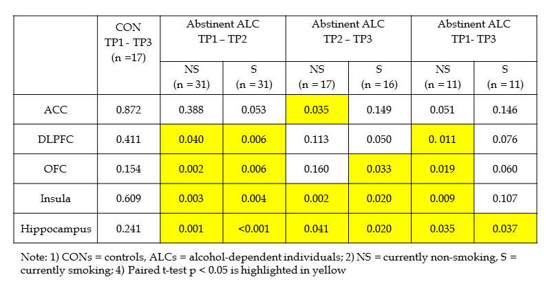
Table 1: Significance levels (p-values of paired t-tests) of regional volume changes between TP1, TP2, and TP3 within CON, and abstinent nsALC and sALC.
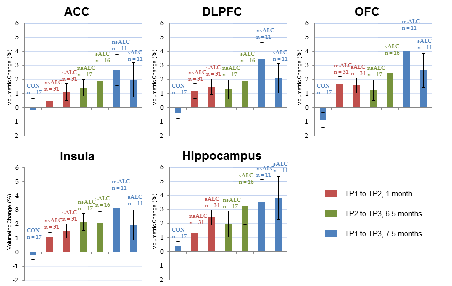
Figure 1: Regional volume changes (percentage of the baseline volume of each pair, mean and standard errors) between TP1, TP2, and TP3 within CON, and abstinent nsALC and sALC.