4227
Altered causal connectivity of the anterior cingulate cortex in obsessive compulsive disorder1Huaxi MR Research Center (HMRRC), Department of Radiology, West China Hospital of Sichuan University, Chengdu, People's Republic of China, 2Department of Psychiatry, West China Hospital of Sichuan University, Chengdu, People's Republic of China
Synopsis
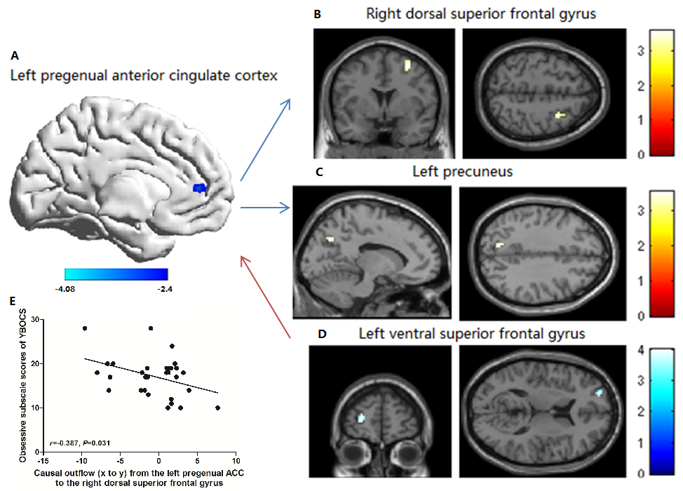
In order to explore the role of anterior cingulate cortex (ACC) in the pathophysiology of obsessive-compulsive disorder (OCD), we used the resting-state functional magnetic resonance imaging (rfMRI) and Granger causality analysis (GCA) and found that the left pregenual ACC of OCD showed decreased amplitude of low-frequency fluctuation (ALFF) than controls. The areas with altered ALFF exhibited decreased driving effect to right dorsal superior frontal gyrus (dSFG) and left precuneus, and a significant increase in the causal influence from left ventral SFG (vSFG) to the left pregenual ACC in OCD compared to controls.
Purpose
Neuroimaging studies of OCD suggested the deficit of the cortico-striato-thalamic-cortico (CSTC) circuits relate to the pathophysiology of OCD.1 The ACC as part of a wider CSTC circuitry contributes to OCD by altered function connectivity. However, previous studies ignored the direction of information flow between brain regions, which is crucial to understand whether the neural function of ACC is primarily abnormal and affects other brain systems or whether it responds normally to an abnormal pattern of information conveyed by other brain structures. Our present aim is to investigate the information flow between the ACC and other brain regions by using rfMRI and GCA.Methods
Thirty-one patients with OCD (aged 27.1±9.5 years) and 36 matched healthy controls (aged 24.6±7.4 years) were recruited. The diagnosis of OCD was made using the Structured Clinical Interview for DSM-IV Axis I Disorders, and the Yale–Brown Obsessive Compulsive Scale (YBOCS) was used to rate the severity of OCD symptoms. The rfMR images sensitized to changes in BOLD signal levels were obtained via a EPI sequence of a 3-Telsa GE MRI system (TR/TE=2000/30msec, flip angle=90°, slice thickness=5mm with no gap, 30 axial slices, 200 volumes in each run).
The DPARSF software was used to perform the data preprocessing and calculate the ALFF that is thought to reflect spontaneous neural activity. We compared the ALFF values of the bilateral ACC between groups, using the bilateral ACC masks from the Automated Anatomical Labeling template.2 Then selecting the cluster with altered ALFF between groups as a seed, we continued to explore if the region had altered directional influence with broader brain circuitry by using the GCA. We performed the first-order voxel-wise coefficient-based GCA by using the REST software,3 which allows for the physiological possibility that bidirectional influences of opposite effects could simultaneously exist in the brain, and the signed-path coefficient maps allow parametric statistical analysis for group-level inference.
The two-sample t test in SPM8 was performed to explore ALFF differences between groups (cluster level family-wise error [FWE] corrected P<0.05). For between-group comparison of GCA, two-sample t tests were used to compare signed-path coefficient maps between patients and controls (cluster level FWE corrected P<0.05), which were restricted to the voxels with excitatory or inhibitory influence from and to the seed region in both groups by using an explicit mask from the union set of one-sample t test results of the two groups (voxel-level uncorrected P<0.001 and cluster-level uncorrected P<0.05), respectively.
Results
We found decreased ALFF in the left pregenual ACC (P=0.029) of OCD than controls (Figure 1A). There’s no significant difference of ALFF in the right ACC between the two groups.
Two-sample t test revealed a significant difference between groups in the causal outflow from the left pregenual ACC to right dSFG (P=0.016) (Figure 1B), where the controls showed an excitatory influence (path coefficient=2.24±3.82), while the patients exhibited an inhibitory influence (path coefficient=-0.88±4.13). In addition, there was a significant difference in the effect of left pregenual ACC on left precuneus (P=0.019) (Figure 1C), where the controls exhibited an excitatory influence (path coefficient=3.23±3.38), while the patients demonstrated an inhibitory influence (path coefficient=-0.21±4.57). Patients also showed a significant increase in the causal influence from left vSFG to the left pregenual ACC (path coefficient=0.42±1.06) compared to controls (path coefficient=-0.68±1.09) (P=0.029) (Figure 1D).
In patients group, the path coefficient from the left pregenual ACC to the right dSFG showed significant correlations with the obsessive subscale scores of YBOCS (r=-0.387, P=0.031) (Figure 1E).
Discussion
The decreased driving effect from left pregenual ACC to right dSFG and left precuneus might be caused by the decreased neural spontaneous function reflected by lower ALFF, suggesting the OCD patients had less excitatory influence from the left pregenual ACC to the regions relevant to cognitive function and default mode network (DMN). Compared with controls, OCD also demonstrated increased feedback, suggesting patients had a less inhibitory to the left pregenual ACC, which might represent a compensatory factor that is meant to protect against the deficit function of ACC.Conclusion
Our present study suggested that patients with OCD had abnormal functional causal connectivity in cognitive and affective brain systems, particularly including lateral frontal and DMN that were involved in the underlying pathology of OCD. The correlation between the GCA value from the left pregenual ACC to the right dSFG and obsessive subscale scores suggested the causal effect may reflect the disease severity of OCD.Acknowledgements
No acknowledgement found.References
1. Menzies L, Chamberlain SR, Laird AR, et al. Integrating evidence from neuroimaging and neuropsychological studies of obsessive-compulsive disorder: the orbitofronto-striatal model revisited. Neurosci Biobehav Rev. 2008;32(3):525-49.
2. Tzourio-Mazoyer N, Landeau B, Papathanassiou D, et al. Automated anatomical labeling of activations in SPM using a macroscopic anatomical parcellation of the MNI MRI single-subject brain. Neuroimage. 2002;15(1):273-89.
3. Zang ZX, Yan CG, Dong ZY, et al. Granger causality analysis implementation on MATLAB: a graphic user interface toolkit for fMRI data processing. J Neurosci Meth. 2012;203(2):418-26.
Figures