4223
Discrimination of patients with first episode schizophrenia via quantitative cortical morphology features and machine learning methodsHuaiqiang Sun1, Ying Chen1, Haoyang Xing1, Su Lui1, and Qiyong Gong1
1Huaxi MR research center, Department of Radiology, West China Hospital, Sichuan University, Chengdu, People's Republic of China
Synopsis
A multivariable analysis framework for schizophrenia prediction with quantitative cortical morphology features features extracted at individual level.
Purpose
Schizophrenia has long been considered a neurodevelopment disease, aberrant cerebral morphology may involve in the pathology of the disease. Extensive works have been done in the aspect of volume, cortical thickness, surface area and shape of certain brain region.1 However, no consistent results have been reached. A potential explanation is that most studies typically report group level differences on limited samples and the robustness of differences at individual level is not well established. The current study extracted shape features at individual level and try to establish a framework to evaluate to importance of extracted features and construct predictive model using selected important features. The performance of predictive model can further reflects the robustness of features exhibited difference between patients and controlsMethods
Fifty first-episode right handed schizophrenia patients (26 male, 24 female, age from 18 to 20, mean age 19.1) recruited from outpatient and inpatient units of the Mental Health Center were included in this study. Confirmation of diagnosis was determined by clinical psychiatrists using the Structured Clinical Interview for DSM-IV (SCID). All the patients did not receive any type of medication before MRI scanning. The illness duration of all patients was less than 2 years. Fifty-four controls (25 male, 29 female, age from 18 to 20, mean age 19.5) were recruited from the local community via poster advertisements. High resolution T1 weighted anatomical images were acquired on a clinical 3T scanner with a 8-channel phase array head coil using 3D spoiled gradient (3D-SPGR) sequence (TR=8.5ms, TE=3.5ms, TI=400ms, Flip angle=12) with 240 x 240 matrix over a field of view of 240 x 240 mm and 156 axial slices of 1mm thickness. T1-weighted anatomical images were processed with both Freesurfer's recon-all processing pipeline with Desikan-Killiany-Tourville (DKT) atlas and the ANTs antsCorticalThickness pipeline to generate labeled cortical and non-cortical volumes. Then the labeled brains were feed into an open source automatic feature extraction toolkit named Mindboggle (www.mindboggle.info). Hybrid gray/white segmentation was created from both FreeSurfer and ANTs output. Surface meshes were generated from each segmented region. The following measures were computed: (1) Volume and surface area of all labeled regions. For each participant, the volume of each labeled region was normalized by his/her total cerebral volume; (2) Surface shape measures (local cortical thickness, curvature, convexity, travel and geodesic depth) for every cortical mesh vertex within each label and sulcus; (3) Statistical properties (mean, standard deviation, skew, kurtosis) for each shape measure in step (2) within each label and sulcus. The all-relevant feature selection (implemented in R package “Boruta”)2 and the construction and evaluation of random forest classifier were performed within same cross-validation loops.3 The overall accuracy, sensitivity/specificity, kappa score were used to characterize the performance of the classifiers. Features that were selected in more iterations than would be expected to occur at random were identified as significant features. The whole workflow was illustrated in Figure 1Results
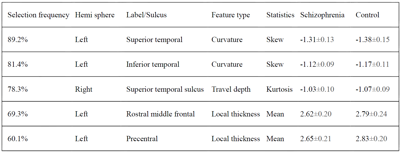
A total of 2941 features represent cerebral shapes were extracted from each participant. In building and evaluation the classifier for discriminating schizophrenia patients and controls, we performed 100 runs of 10-fold stratified cross-validation, and computed the mean classifier performance from a total of 1000 folds. The mean classification accuracy and kappa value achieved via repeated 10-fold cross-validation is 78.7% and 0.61, using features selected by all-relevant feature selection algorithm. The mean sensitivity and specificity for schizophrenia patients is 74.2% and 83.0%, respectively. As we embedded the feature selection in cross-validation procedure, thus a total of 1000 feature subsets were obtained in each run of classifier construction. The mean length of obtained feature subsets is 9.3 (range from 6 to 12, from 0.2%–0.4% of all extracted features). Significant features were list in Table 1.Discussion
The current result confirmed the aberrant cortical morphology in temporal and frontal lobe of patients with first episode schizophrenia at individual level. In addition, the current results further refined previous voxel based morphometry (VBM) studies that report reduced gray matter in patients with schizophrenia as gray matter volume measured in VBM can be sensitive to changes in gray matter volumes as well as differences in cortical surface curvature, which cannot be distinguished from each other.Conclusion
The current study established a multivariable analysis framework for schizophrenia prediction with structural features extracted at individual level. The proposed framework has the ability to figure out the features that relevant to classification, which can be used as imaging biomarkers and help to understand the underlying mechanism of the disease. In addition, the selection frequency and classifier performance reflect the robustness of the selected imaging markers.Acknowledgements
No acknowledgement found.References
1. Vita a, De Peri L, Silenzi C, Dieci M. Brain morphology in first-episode schizophrenia: a meta-analysis of quantitative magnetic resonance imaging studies. Schizophr Res. 2006;82(1):75–88.
2. Kursa MB, Rudnicki WR. Feature Selection with the Boruta Package. J Stat Softw. 2010;36(11):1–13.
3. Smialowski P, Frishman D, Kramer S. Pitfalls of supervised feature selection. Bioinformatics. 2009;26(3):440–443.
Figures
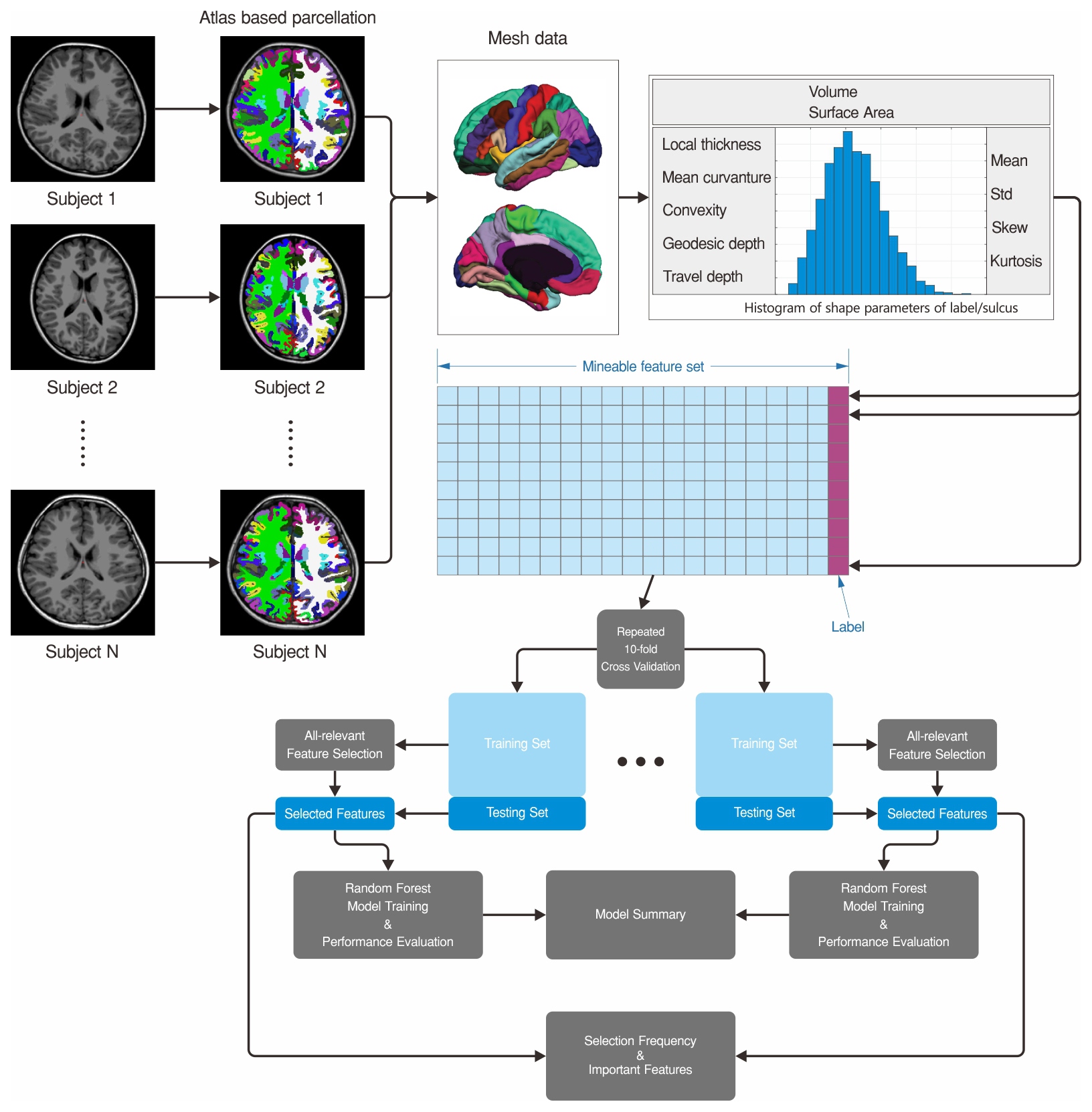
Figure 1. The whole workflow of feature extraction, selection and classifier training.
Table
1 Significant features for discriminating patients
with first episode schizophrenia versus healthy controls