4222
Resting-state functional connectivity abnormalities associated with posttraumatic stress disorder and traumatic stress: a graph-based analysis1Department of Radiology, Huaxi MR Research Center, Chengdu, People's Republic of China
Synopsis
Previous studies have clarified that differentiating the disease- and traumatic stress-related brain changes may elucidate the core neural mechanism of PTSD. This study is to investigate brain functional alterations in PTSD and the traumatic exposed controls (TEC) relative to the non-traumatized healthy controls (HC) separately, using a data-driven graph theoretical approach--whole-brain functional connectivity strength (FCS) mapping. The current study provided the preliminary evidence of common and separate abnormalities of neural correlates at whole-brain level associated with PTSD and traumatic stress. The disequilibrium between the DMN and the SN might be associated with the pathophysiology of PTSD.
Backgroud
Posttraumatic stress disorder (PTSD) is a trauma- or stressor-related disorder that is characterized by four clusters of symptoms: reexperience, hyperarousal, avoidance and negative cognition and mood. In recent decades, many neuroimaging studies have indentified brain functional alterations in PTSD. However, due to differences in the selection of control subjects, it is difficult to conclude whether the observed alterations were related to disease or traumatic stress. Previous voxel-based meta-analysis of gray matter has clarified that differentiating the disease- and traumatic stress-related brain changes may elucidate the core neural mechanism of PTSD [1]. The purpose of this study is to investigate brain functional alterations in PTSD and the traumatic exposed controls (TEC) relative to the non-traumatized healthy controls (HC) separately, using a data-driven graph theoretical approach--whole-brain functional connectivity strength (FCS) mapping [2], and evaluate the relationship between altered cerebral FCS and symptom severity in PTSD.Methods
Data acquisition was conducted on a 3T MR imaging system (EXCITE, General Electric, USA) from right-handed earthquake survivors, including 93 drug-naïve first episode PTSD patients, 88 TEC within 7-15 months after the earthquake, as well as 99 HC. All images were preprocessed using SPM8 and DPARSF software including slice timing, head-motion correction, and normalization to Montreal Neurological Institute (MNI) space. Then we subjected preprocessed functional runs to voxel-based whole brain correlation analysis, resulting in a Pearson correlation coefficient matrix, and the FCS of the given voxel were computed after the FCS maps were spatially smoothed with a 6mm Gaussian kernel using SPM8. The FCS maps were compared between two groups of the PTSD patients, TEC and HC by using the voxel-based two-sample t-test in SPM8. Partial pearson correlation analyses were performed to identify the association between altered neural correlates and symptom severity as measured by clinician-administered PTSD scale (CAPS) and PTSD checklist (PCL) scores with the age as a covariate.Results
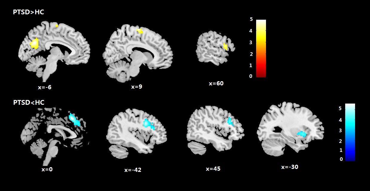
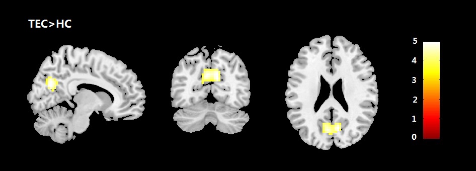
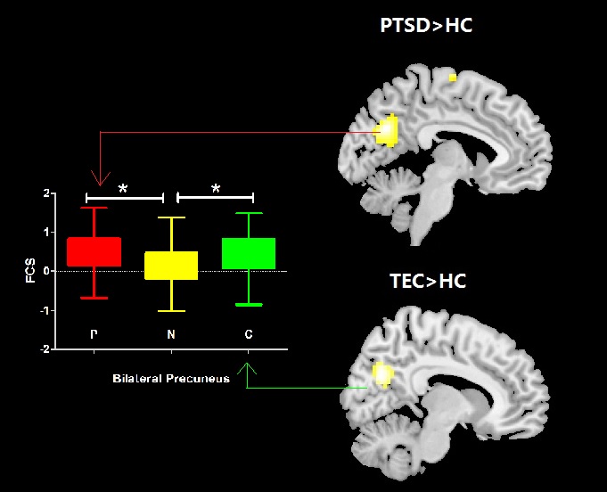
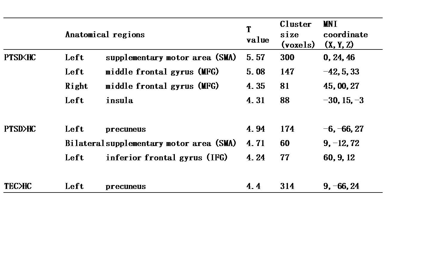
Compared to HC, PTSD patients showed increased FCS in the left precuneus, bilateral supplementary motor area (SMA), and right inferior frontal gyrus (IFG), and decreased FCS in the left insula, and bilateral middle frontal gyrus (MFG) and SMA (P <0.05, Family-wise error correction ) (Fig.1). Moreover, compared with HC, TEC showed overlapping increased FCS in the left precuneus with PTSD patients (P <0.05, Family-wise error correction ) (Fig.2 and 3) (Table 1). There were no significant FCS alterations between PTSD and TEC. Correlation analysis in PTSD patients revealed no significant associations between the FCS in abnormal brain regions and the CAPS score.Discussion
Both PTSD and TEC showed increased FCS in the left precuneus, a region suggested to be the 'core node' or 'hub' of the default mode network (DMN) that is activated during "resting consciousness". This may indicate a common functional impairment in self consciousness associated both with PTSD and traumatic stress. Disrupted nodal properties of the functional connectome in the DMN including the precuneus were also reported in a recent graph theoretic study in PTSD [3]. Interestingly, more regions were identified only in PTSD with increased FCS in the bilateral SMA, and right IFG, and decreased FCS in the left insula, bilateral MFG and SMA. We speculated this phenomenon may indicate unique functional alterations in PTSD that a greater need for the recruitment of emotion and cognition control. The SMA located in the dorsomedial frontal cortex, and the MFG and IFG located in the dorsolateral frontal cortex, responding for cognitive management. This is consistent with a previous PTSD study reported greater blood oxygenation level dependent (BOLD) signals in the dorsomedial frontal cortex in response to emotional relative to neutral stimuli. The insular cortex mainly located in the front part, which is a key node of the salience network (SN). The SN is responsible for detecting and responding to salient stimuli, and help to maintain an adaptive balance between internal mentation and externally-oriented focus. There is evidence that disequilibrium between SN and DMN was reported in PTSD.Conclusion
The current study provided the preliminary evidence of common and separate abnormalities of neural correlates at whole-brain level associated with PTSD and traumatic stress. The disequilibrium between the DMN and the SN might be associated with the pathophysiology of PTSD.Acknowledgements
This study was supported by the National Natural Science Foundation(Grant Nos. 81220108013, 81227002 and 81501452), and Programfor Changjiang Scholars and Innovative, Research Team in University(PCSIRT, Grant No. IRT1272)of China. Dr. Gong would also like toacknowledge the support from his Changjiang Scholar ProfessorshipAward (Award No. T2014190) of China and the CMB DistinguishedProfessorship Award (Award No. F510000/G16916411) administeredby the Institute of International Education, USA.References
[1] Li L, Wu M, Liao Y, Ouyang L, Du M, Lei D, Chen L, Yao L, Huang X, Gong Q. Grey matter reduction associated with posttraumatic stress disorder and traumatic stress. Neuroscience & Biobehavioral Reviews. 2014 Jun;43:163-172.
[2] Hou JM, Zhao M, Zhang W, Song LH et al., Resting-state functional connectivity abnormalities in patients with obsessive-compulsive disorder and their healthy first-degree relatives. J Psychiatry Neurosci. 2014 Sep;39(5):304-11.
[3]Lei D1, Li K1, Li L1, Chen F et al., Disrupted Functional Brain Connectome in Patients with Posttraumatic Stress Disorder.Radiology.2015 Sep;276(3):818-27.
Figures