4214
Substructural Volumes of the Thalamus in Alcoholism1Psychiatry and Behavioral Sciences, Stanford University, Stanford, CA, United States, 2Neurosciences, SRI International, Menlo Park, CA, United States, 3Medical Imaging, Univ. of Arizona, Tucson, AZ, United States
Synopsis
Volumes of the thalamus and 4 thalamic substructures (i.e., anterior (AT), mediodorsal (MD), ventrolateral (VL), pulvinar (Pul)) were quantified using a novel automated segmentation algorithm in 18 individuals meeting criteria for Alcohol Use Disorders (AUD) and 28 healthy controls (Con). Multiple regressions considered contributions of diagnosis (i.e., Con vs. AUD), age, sex, and intracranial volume on substructural volumes. Volumes of AT, VL, and Pul were smaller with increasing age. Volumes of MD and Pul were affected by diagnosis: both were smaller in the AUD relative to the Con group. These results suggest that thalamic substructures have differential vulnerability to pathological processes.
Introduction
The current standard of measuring global, undifferentiated brain structures is too coarse to distinguish substructural volumes, such as those of the thalamus, which may be differentially affected by Alcohol Use Disorders (AUD) and other pathologies. Recently, a white-matter-nulled MPRAGE sequence [1] for manual [2] or automatic [3] segmentation of thalamic nuclei has shown considerable promise. We report here, for the first time, the use of a fast, automated thalamic segmentation algorithm to investigate the effects of AUD on thalamic substructures.Materials and Methods
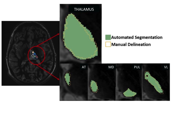
After informed consent, 46 human subjects (28 healthy, controls aged 56.14±12.23, 14 women; 18 meeting criteria for AUD aged 52.84±7.41, 6 women) were scanned on a GE 3T MRI scanner using an 8-channel head array coil. A white-matter-nulled MPRAGE sequence [1] with 1mm isotropic spatial resolution was acquired. A multi-atlas comprising 20 prior white-matter-nulled MPRAGE datasets (12 patients and 8 controls) was used for automatic segmentation. This atlas was constructed based on manual delineations of thalamic substructures performed by a neuroradiologist guided by the Morel atlas [2] (Fig 1). The template and input images were cropped to include just the bilateral thalami, which accelerated the processing time and minimized errors due to whole brain registration. Four subregions of the thalamus were considered: anterior (AT), mediodorsal (MD), ventrolateral (VL), pulvinar (Pul). Analysis. A multiple regression on total thalamic volume (Thal) included 4 factors: intracranial volume (ICV), Diagnosis, Age, and Sex. Multiple regressions for each substructural volume included 5 factors: Thal, ICV, Diagnosis, Age, and Sex.Results
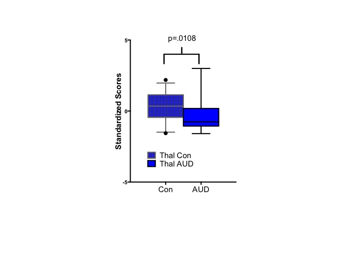
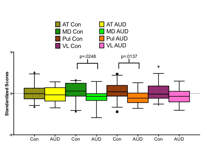
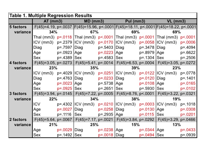
Diagnosis, Age, and Sex together explained 40% of the variance in Thal (smaller in AUD, women, and with increasing age): ICV did not significantly modulate Thal. Table 1 presents the results of multiple regressions for each of the 4 substructures, starting with 5 factors. Thal explained a significant percent of the variance of each sub-region. Together, the remaining 4 factors explained 23–39% of the variance of substructural volumes. Volumes of AT and VL were most sensitive to Age (smaller with increasing age). MD was sensitive to Diagnosis and Sex (smaller in AUD and in women). Pul was similarly sensitive to Diagnosis and also to Age (smaller in AUD and with increasing age). Indeed, when volume measures were corrected for Age, Sex, and ICV, Thal (Fig 2), MD, and Pul (Fig 3) were found to be significantly smaller in the AUD relative to the control group.Discussion
These results demonstrate the utility of using automatic segmentation of 3T MPRAGE data to delineate substructures of the thalamus. Total thalamic volume (i.e., Thal) was sensitive to Diagnosis, Age, and Sex. Although volumes of all 4 thalamic substructures correlated significantly with Thal, older age was associated with smaller volumes of the AT, Pul, and VL. MD was selectively affected by Diagnosis, a finding that comports a previous study conducted on postmortem tissue showing that MD is selectively sensitive to AUD [3]. Future work will evaluate how additional factors, such as drinking variables, hematological indices, and cognitive performance are related to substructural volumes of the thalamus to provide further support for the biological relevance of these quantified volumes.Acknowledgements
NIH Grants: U01 AA017347, R01 AA005965, R21 AA023582References
[1] Saranathan, M., et al., Optimization of white-matter-nulled magnetization prepared rapid gradient echo (MP-RAGE) imaging. Magn Reson Med, 2015. 73: 1786-1794.
[2] Tourdias, T., et al., Visualization of intra-thalamic nuclei with optimized white-matter-nulled MPRAGE at 7T. Neuroimage, 2014. 84: 534-545.
[3] Su et al., Proceedings of ISMRM 2015. p6231.
[4] Harding, A., et al., Degeneration of anterior thalamic nuclei differentiates alcoholics with amnesia. Brain, 2000. 123: 141-154.
Figures