4212
White matter structure asymmetry of drug-naïve first-episode schizophrenia patients under different genetic load1West China Hospital, Chengdu, People's Republic of China
Synopsis
We analyzed the white matter (WM) asymmetry in a relatively large sample of drug-naïve schizophrenia patients under different genetic load. Based on this method, our findings demonstrated similar overall WM brain torque in sporadic and familial schizophrenia patients and healthy controls. Furthermore, our observation that familial illness was associated with more abnormal brain asymmetry compared to sporadic patients and that these specific changes were mainly located in a functional network associated with auditory hallucinations suggesting genetic factors may play critical roles in impacting risk for the most common form of hallucinations in schizophrenia.
Propose
Hemispheric asymmetry has been regarded as a functional trait, especially related to language functions1. Rightward frontal, leftward occipital and the planum temperale could be seen among most of the healthy adults2. This property might be different in the schizophrenia patients. Schizophrenia has been regarded as a disorder of the genetic mechanisms that control the development of cerebral asymmetry3. Changes of asymmetry pattern of brain structure have been reported, including reduction or inversion in schizophrenia patients under different genetic load. However, studies concerning abnormal asymmetry of white matter (WM) in schizophrenia have been inconsistent. The aim of current study was to confirm an abnormal asymmetry of WM in patients with schizophrenia and to determine whether these alterations were differentially expressed in familial vs. sporadic schizophrenia.Methods
All first episode drug-naïve patients (n=72) met criteria for schizophrenia based on the Structured Clinical Interview for DSM-IV Axis I Disorder-Patient Edition. They were divided into a ‘familial’ group (n=42) having a positive family history (FH+) of schizophrenia and a ‘sporadic’ group (n=30) (FH-) by clinical interview according to the Murray’s definition. 72 healthy individuals matched for gender, age, education level with the schizophrenia group were included. All magnetic resonance imaging scans were performed on a 3.0T MRI scanner (EXCITE, General Electric, Milwaukee, USA). High resolution T1-weighted images and DTI data were acquired by 3D-SPGR and single-shot spin-echo echo-planar imaging sequence separately. Lateralization index (LI) of white matter and gray matter volume as well as fractional anisotropy and mean diffusion of white matter were calculated. The calculation were based on the following formula4: LI = 2(original image – flipped image) ⁄ (original image + flipped image) Hemispheric level, voxel-wise and correlation analyses were then done based on these parameters.Results
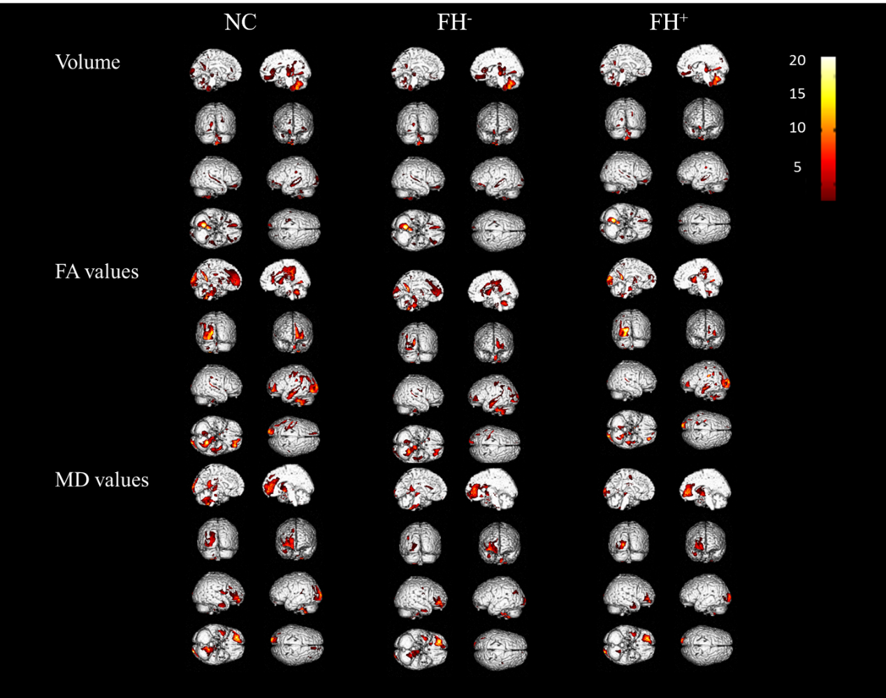
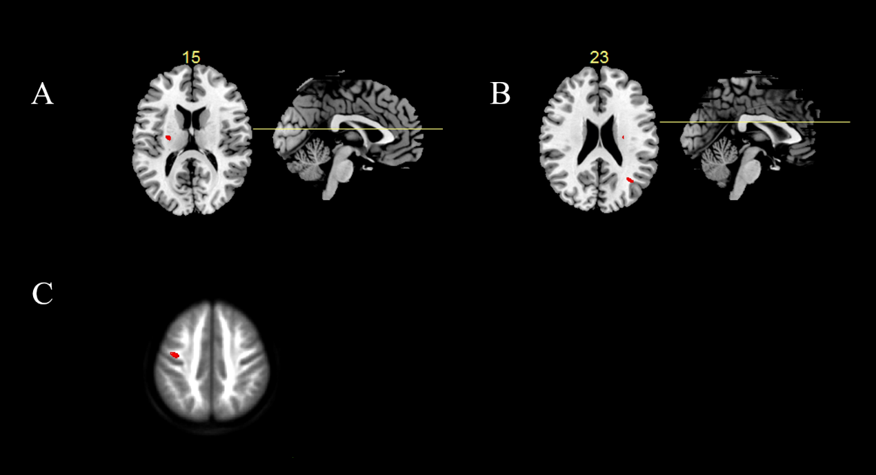
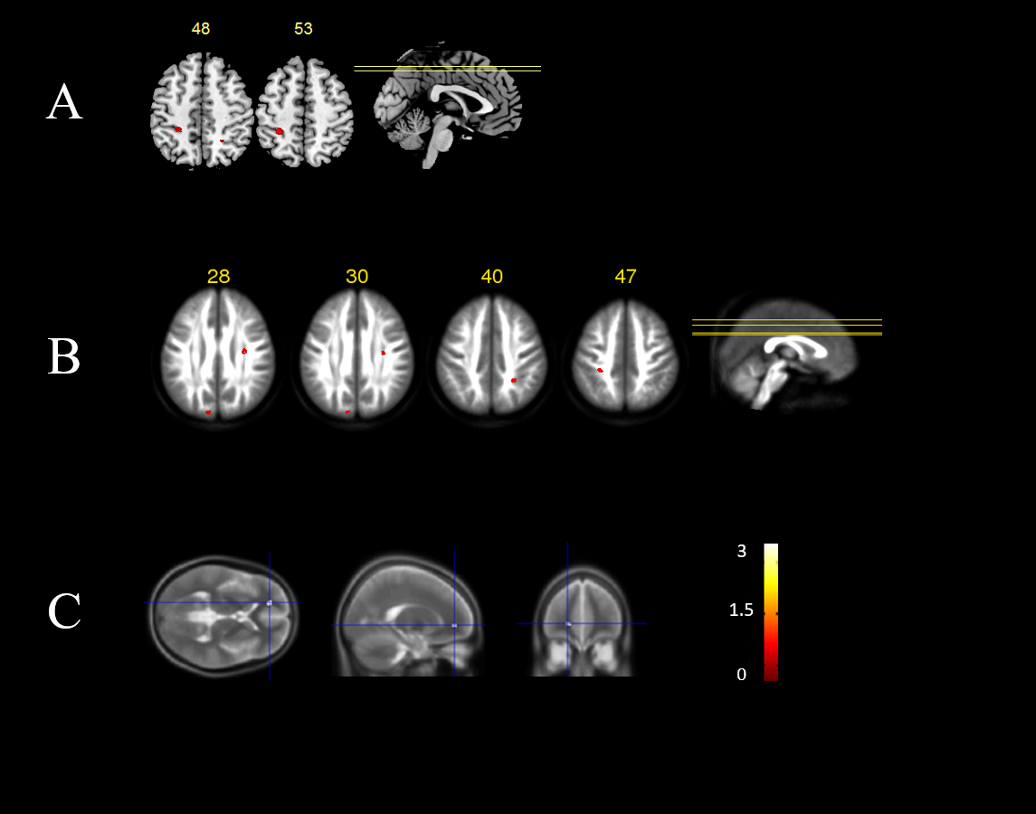
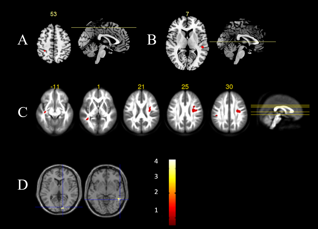
In the volume study, all 3 groups showed similar overall rightward volume brain torque at the hemisphere level. The comparison of the LI of left and right hemisphere found that NC and FH- group showed significant differences while there was no significance difference detected in the FH+ group (Figure 1). Compared with NC, sporadic patients showed reversed leftward lateralization of WM volume in posterior limb of internal capsule volume and increased rightward asymmetry in the posterior corona radiate, as well as reverse of leftward lateralization of FA value of WM in postcentral gyrus. There was no abnormal change in asymmetry of MD value in sporadic patients (Figure 2). There were more areas of asymmetry alterations found in the familial patients: they showed (1) reversed leftward lateralization of WM volume in inferior parietal lobule (IPL) (2) reversed leftward lateralization of FA value of WM in cuneus (3) reversed rightward lateralization of volume in superior parietal lobule (4) reversed rightward lateralization of FA value of anterior corona radiate and WM in IPL (5) decreased leftward lateralization of FA value in WM of postcentral gyrus and (6) increased MD value of WM in anterior cingulate gyrus (ACC) (Figure 3). Compared with sporadic patients, familial patients had (1) decreased leftward lateralization of volume in IPL (2) increased rightward asymmetry of volume in superior temporal gyrus (3) increased leftward asymmetry of FA value in temporal stem, WM of middle temporal gyrus and supramarginal gyrus (4) increased leftward lateralization in MD value of WM in lingual gyrus and middle temporal gyrus and (5) reversed rightward lateralization of FA value in anterior corona radiate and WM of precentral gyrus (Figure 4). Greater laterality alterations of FA in right middle temporal gyrus correlated negatively with clinical symptoms.Discussion & Conclusion
Compared with the sporadic patients, familial
patients showed greater WM asymmetry abnormalities related to auditory cortex
and a posterior language network. Greater laterality alterations of FA in right
middle temporal gyrus correlated negatively with clinical symptoms. Our
observation that familial illness was associated with more abnormal brain
asymmetry compared to sporadic patients and that these specific changes were
mainly located in a functional network associated with auditory hallucinations
suggests that familial and perhaps genetic factors may impact brain anatomy in
ways that impact risk for the most common form of hallucinations in
schizophrenia.Acknowledgements
This study was supported by the National Natural Science Foundation (Grant Nos. 81601458, 81621003, 81220108013, 81227002 and 81030027), National Key Technologies R&D Program (Program No. 2012BAI01B03) and Program for Changjiang Scholars and Innovative Research Team in University (PCSIRT, Grant No. IRT1272) of China. Dr. Gong would also like to acknowledge the support from his Changjiang Scholar Professorship Award (Award No. T2014190) of China and American CMB Distinguished Professorship Award (Award No. F510000/ G16916411) administered by the Institute of International Education, USA.References
1. Hervé P Y et al. Trends in ognitive sciences, 2013, 17(2): 69-80.
2. Park H J et al. NeuroImage, 23:213-23.
3. Crow T J et al. Brain research reviews, 31:118-29.
4. Kawasaki Y et al. Biological psychiatry, 63:793-800.
Figures