4210
Modular organization of functional connectivity in schizophrenia patients beyond the resolution limit1Center for Neuroscience and Cognitive Systems, Istituto Italiano di Tecnologia, Rovereto (TN), Italy
Synopsis
Graph theoretical methods have been widely applied to study the modular organization of functional connectivity networks in neuropsychiatric disorders like Schizophrenia. However, current methods are affected by a resolution limit that prevents detection of modules that are smaller than a scale determined by the size of the entire network. We have developed a resolution-limit-free method, dubbed Surprise, and applied it to study resting state functional connectivity networks in a large cohort of Schizophrenia patients and matched controls. Improved resolution reveals substantial reorganization of resting state connectivity structure in patients, with previously undetected fragmentation and merging of sensory and associative modules.
Introduction
Abnormal patterns of brain functional connectivity have been observed in a number of neuropsyhiatric conditions, including schizophrenia (SZ). The effects of these alterations on the modular organization of brain connectivity networks can be investigated using a graph representation of the resting state fMRI data and analytical methods derived from graph theory1. Previous results using standard community detection methods have shown reduction in functional connectivity strength in SZ patients, with a largely preserved modular organization. However, it has been recognized that current methods suffer from a resolution limit as they fail to detect modules that are smaller than a scale determined by the size of the entire network. This fundamental limitation may have affected previous studies, and prevented detection of differences in the modular structure of connectivity networks in SZ patients and controls. Recently, we have developed a resolution-limit-free method grounded on probability theory that overcomes these limitations2,3. Here, we present the first application to a large dataset of resting state fMRI data from SZ patients vs. healthy controls.Methods
This study used the resting state fMRI sample data of 78 schizophrenia (strict) patients and 91 healthy control subjects from the Center for Biomedical Research Excellence (COBRE)4 ( COINS:http://coinsmrn.org/dx). The nodes of the connectivity networks were defined from a brain atlas parcellation scheme comprising 638 regions5. The edge weights were calculated from the inter-regional Pearson correlation coefficients. Group-average networks were computed for the two cohorts by Fisher-transformation and averaging of the individuals’ connectivity matrices. Asymptotical Surprise, a fitness function that has been shown to resolve the modular structure of connectivity graphs with unprecedented resolution and accuracy2,3, was used to extract the modular structure of the resting state networks. Network Based Statistics was used to compare connectivity in the two groups, and network- and node-wise parameters, including global efficiency and participation coefficients, were calculated for the two groups.Results
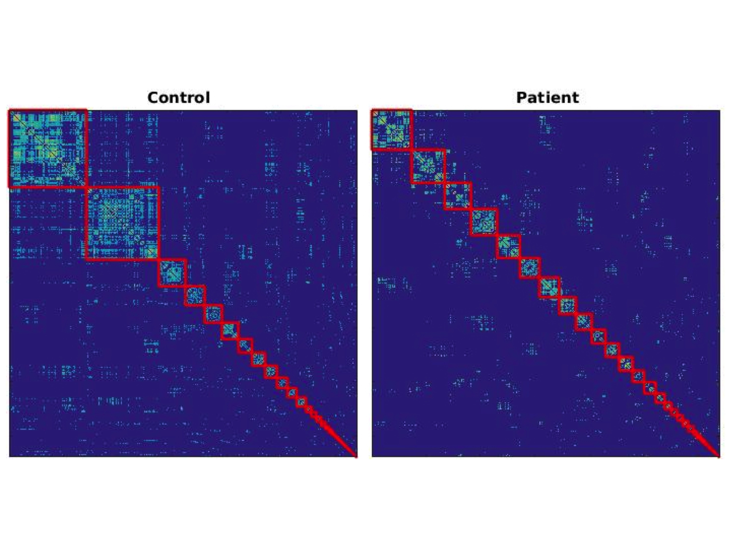
The patients’ group showed overall weaker resting state functional connectivity compared to controls, with a significant decrease in average degree (25.73 vs 53.83, respectively) and global efficiency (0.23 versus 0.32, respectively), in line with previous studies. Surprise maximization resulted in a finer subdivision of modules than previously reported, with a heterogeneous size distribution and a comparable number of modules in the two groups (41 and 39 in controls and patients, respectively) (Fig. 1). However, the modular organization of resting state networks appears substantially different in the two cohorts (Fig. 2). Notable differences include the fragmentation of the controls’ large motorsensory module into three smaller modules, which however include parts of the associative parietal cortices that are not associated with sensory networks in the control group. Similarly, connectivity of the temporal and supramarginal cortices appears substantially reorganized in SZ patients, with language and auditory structures mixing into functionally integrated modules.Discussion
The observation of a comparable number of modules in the two cohorts is somewhat surprising in the light of the strong reduction in overall connectivity in the SZ group. The disintegration of the sensory module in patients, and the mixing of sensory and associative modules may account for some of positive symptoms of SZ, which include sensory hallucinations. The reorganization and integration of auditory and language modules is particularly interesting, and may provide a mechanism for the auditory hallucinations often experienced by SZ patients.Conclusion
Optimization of Surprise made it possible to study the modular organization of functional connectivity beyond the resolution limit that characterizes other widely applied graph theoretical methods. In sharp contrast with previous studies, this approach revealed a substantial reorganization of functional modules in patients that may provide a key to understand some of the symptoms of this complex disease.Acknowledgements
No acknowledgement found.References
1. Bullmore, E. T. & Sporns, O. Complex brain networks: graph theoretical analysis of structural and functional systems. Nat. Rev. Neurosci. 10, 186–198 (2009).
2. Nicolini, C. & Bifone, A. Modular structure of brain functional networks: breaking the resolution limit by Surprise. Sci. Rep. 19250 (2016).
3. Nicolini, C., Bordier, C. & Bifone, A. Modular organization of weighted brain networks beyond the resolution limit. NeuroImage (in press).
4. Çetin, M. S. et al. Thalamus and posterior temporal lobe show greater inter-network connectivity at rest and across sensory paradigms in schizophrenia. Neuroimage 97, 117–126 (2014).
5. Crossley, N. A. et al. Cognitive relevance of the community structure of the human brain functional coactivation network. Proc. Natl. Acad. Sci. 110, 11583–11588 (2013).
Figures
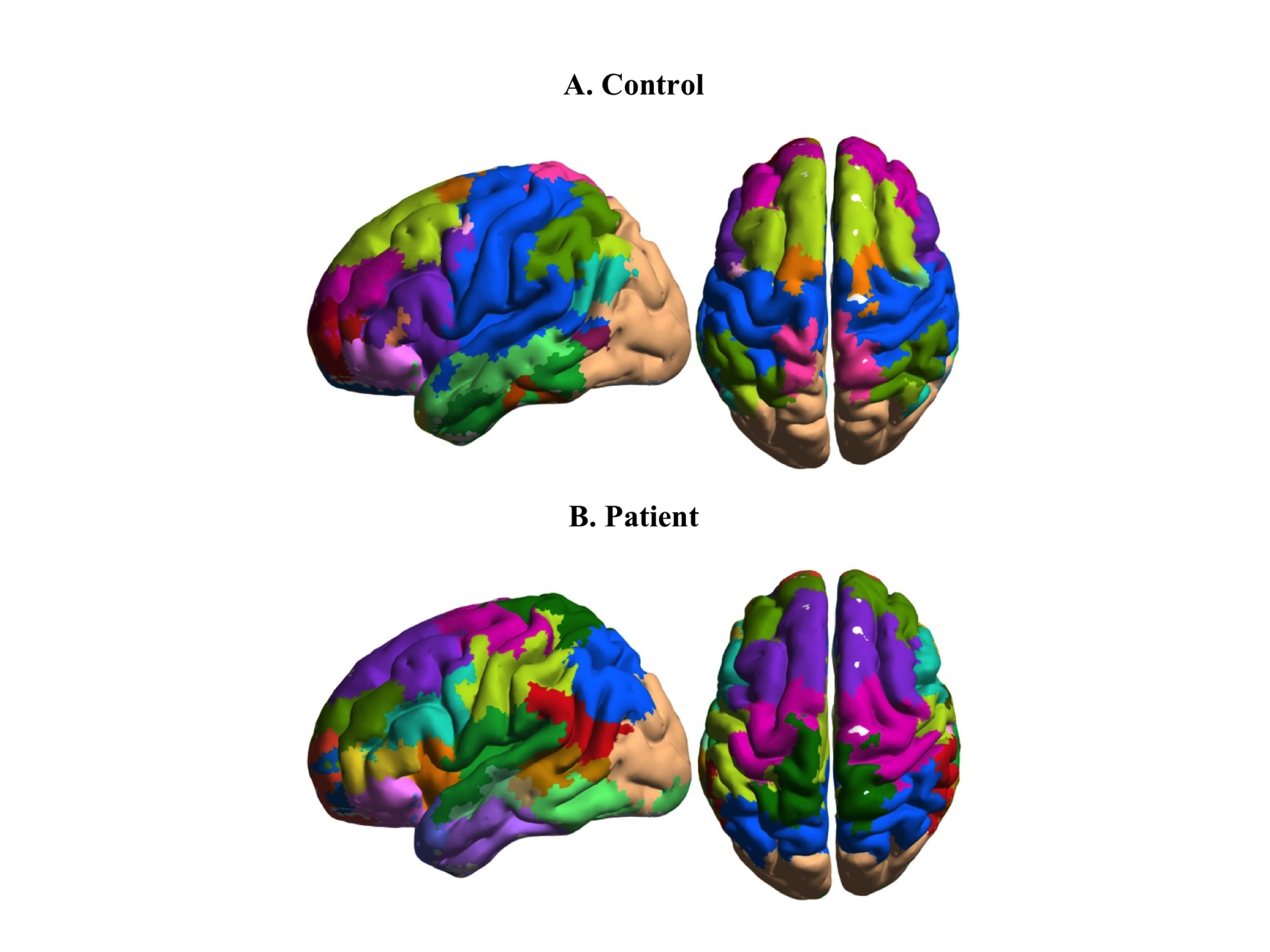
Cortical modules of resting state functional connectivity in the control and SZ groups overlaid on an MRI brain template. Fragmentation and reorganization of sensory and temporal modules are apparent in the SZ cohort. The colors denoting the modules were chosen independently in the two groups to maximize contrast between adjacent structures.