4206
Imaging Mimics of Brain Tumors: Radiologic-Histopathologic Correlation1Department of Radiology, Case Western Reserve University School of Medicine, Cleveland, OH, United States, 2University Hospitals Cleveland Medical Center, Cleveland, OH, United States, 3Department of Pathology, Case Western Reserve University School of Medicine, Cleveland, OH, United States, 4Department of Neurological Surgery, Case Western Reserve University School of Medicine, Cleveland, OH, United States
Synopsis
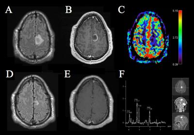
Neuroimaging plays critical diagnostic and prognostic role in the management of brain tumors. However, there can be a significant overlap in the imaging features between neoplastic and various non-neoplastic pathological processes in the brain. The knowledge of key differentiating features of such non-neoplastic entities is essential for a radiologist to facilitate accurate and timely diagnosis. Additionally, in such settings, judicious use of advanced imaging techniques such as perfusion imaging and spectroscopy can help the radiologist as problem-solving tools. In this exhibit, we review the conventional and advanced imaging features of different disease processes that are encountered in day-to-day clinical practice and can mimic brain neoplasms on imaging.
Background
A broad spectrum of non-neoplastic conditions may present with radiological features that are similar to brain tumors. Clues in the initial patient presentation, clinical history, physical examination and laboratory markers can help to a certain extent in differentiating non-neoplastic from neoplastic lesions. However, not uncommonly, these patients may undergo aggressive interventions with significant side effects, only to receive a final diagnosis of a benign pathology. The radiologist can play an important role in such settings by guiding the team towards an appropriate diagnosis and avoiding unnecessary interventions.Purpose
1. To present an overview of different inflammatory, infectious, autoimmune, vascular, iatrogenic and developmental diseases that can present with tumor-like lesions on MR imaging and discuss their key radiological and pathological features. 2. To evaluate the role of advanced imaging techniques such as perfusion, spectroscopy and diffusion imaging in differentiating between neoplastic and various non-neoplastic entities. 3. To formulate an imaging approach for evaluation of tumor-like brain lesions based on conventional and advanced imaging features.
Outline of Content
In this image-rich educational exhibit, a detailed imaging review of common and not so common disease processes that can mimic brain neoplasms is presented. The imaging review is based on conventional as well as advanced imaging techniques. The authors will discuss key pathological features of these disease entities to enhance comprehension of underlying disease processes and thereby facilitate radiological-pathological correlation. Finally, a diagnostic approach for radiological evaluation of tumor mimics is outlined with emphasis on importance of lesion location, conventional imaging characteristics, and spectroscopy and perfusion features.Conclusion
The authors present an interactive educational exhibit on differentiating between tumor-like lesions and brain tumors. This exhibit focuses on detailed imaging review and radiological-pathological correlation of various tumor-like entities and concludes with a practical diagnostic approach for imaging assessment that can be implemented in the day-to-day clinical practice. This exhibit will serve as an engaging self-learning tool for the viewers interested in neuroimaging. Residents and fellows will have an opportunity to familiarize themselves with this recurring and important clinical problem. Practicing radiologists will reinforce their confidence on this topic with this refresher module.Acknowledgements
No acknowledgement found.References
No reference found.Figures
