4203
Imaging features of myoepithelial carcinoma in the nasopharynx and paranasal sinus1MRI Division, the First Affiliated Hostipal of Zhengzhou University, Zhengzhou,China, People's Republic of China, 2Siemens Healthcare Ltd., People's Republic of China
Synopsis
This study aimed to explore the diagnostic points of myoepithelial carcinoma(MEC)in the nasopharynx analyze through analysing the imaging features.11 patients with MEC in the nasopharynx and paranasal sinus confirmed by pathology were analyzed retrospectively. CT and MRI appearances can localize the tumors,show tumors’size, and delineate the relationship of the 1esions with the surrounding tissue.On CT findings, MEC easily has osteolytic destruction and on MR and ADC vuale it owns certain characteristic features.These characteristics are conducive to the early diagnosis and rational treatment in clinics.
Purpose
To analyze the imaging features of myoepithelial carcinoma(MEC)in the nasopharynx and paranasal sinus and explore its diagnostic points.Methods
Data were collected on a MAGNETOM Skyra 3T MR scanner (Siemens Healthcare, Erlangen, Germany) with a 20-channel head coil and a Discovery CT 750 HD. DWI was performed with echo planar imaging technique with the following parameters: TR/TE = 3200/70 ms, FOV = 240 × 240 mm2, slice thickness = 5 mm, interlayer spacing= 1 mm ,19 slices,b value = 0 and 1000 s/mm2. T1WI and T2WI was performed with fast spin echo sequences with the following parameters: TR/TE = 2000/9 ms (T1WI) , TR/TE = 5000/117 ms (T2WI) , FOV = 240 × 240 mm2, slice thickness = 5 mm, interlayer spacing= 1 mm, 20 slices. 11 patients with MEC in the nasopharynx and paranasal sinus confirmed by pathology were analyzed retrospectively. 4 patients underwent CT plain scan,routine MR scan,DWI and DCE-MRI.3 patients underwent routine MR scan,DWI and DCE-MRI.4 patients underwent CT scan.Results
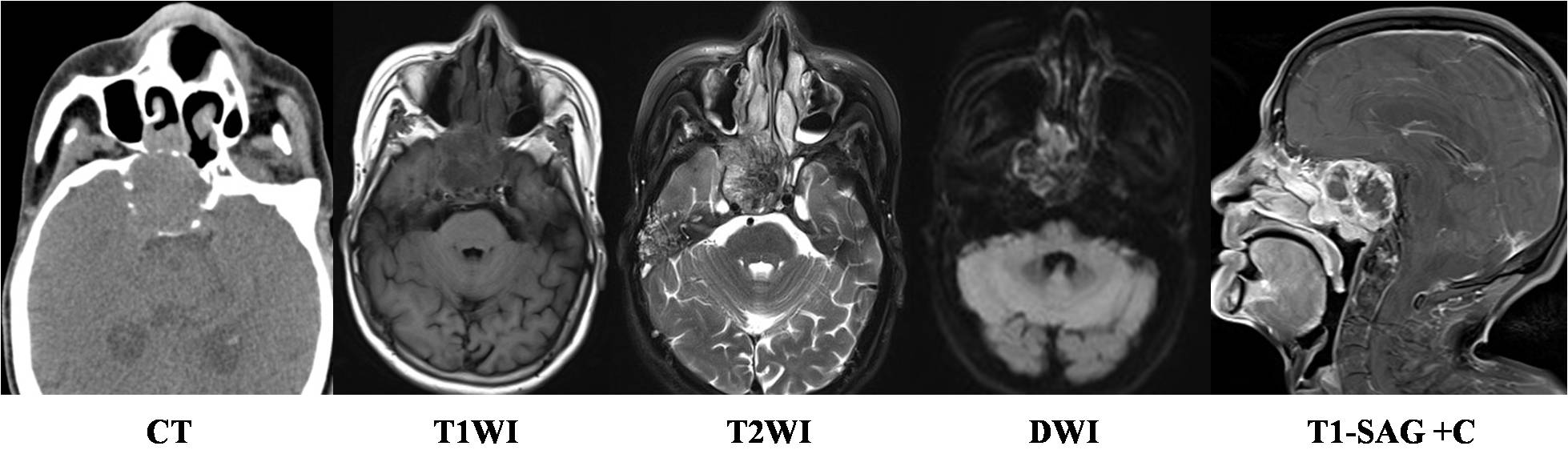
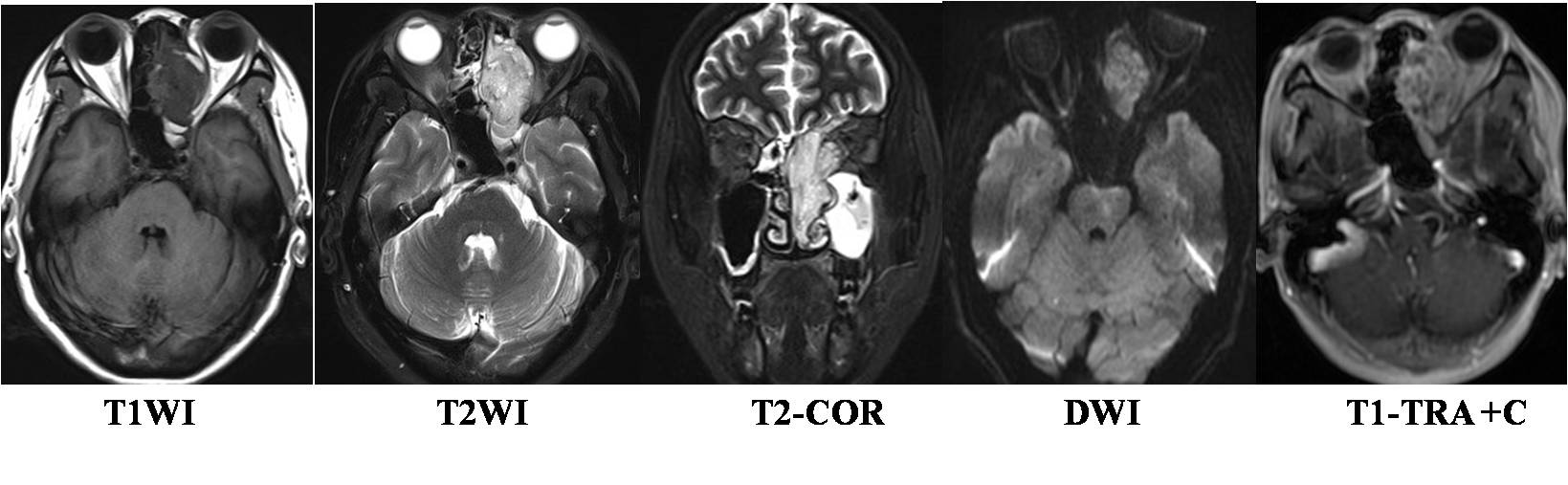
11 cases were one bilateral, irregular shape and unclear boundary.The diameter of the tumors was larger than 2.3 cm. The aggressive nature of the tumors was demonstrated by bone destruction and invasion of adjacent structures. Among 8 patients under going CT examination,tumor centers were located in nasopharynx in 4 cases,3 in maxillary sinus,1 in nasal cavity and ethmoid sinus.8 cases were soft tissue density.5 lesions demonstrated destruction of the adjacent bone structure.2 cases had cervical lymph node metastasis.1 lesion showed slight enhancement. Among 8 patients under going MRI examination, tumor centers were located in nasopharynx in 5 cases,1 in maxillary sinus and 1 in ethmoid sinus.On T1WI,4 showed equal signal and 3 showed low signal.On T2WI,7 showed sligthly high or high signal. After contrast agent injection, 6 patients showed obvious heterogeneous enhancement,1 showed obvious homogeneous enhancement and 1 with circular enhancement of cervical lymph node. The mean ADC value was(0.87±0.04)×10-3mm2/s. The mean ADC value of MEC in the nasopharynx and paranasal sinus was lower than squamous cell carcinoma and adenoid cystic carcinoma and higher than lymphoma in the nasopharynx and paranasal sinus.Conclusion
CT and MRI appearances can show tumors’size,localize the tumors,and delineate the relationship of the 1esions with the surrounding tissue.MEC in the nasopharynx and paranasal sinus easily has osteolytic destruction on CT and owns certain characteristic features on MR and ADC vuale. These features are helpful to the clinical diagnosis and the treatment plan formulation.Acknowledgements
No acknowledgement found.References
[1] Soon G, Petersson F. Myoepithelial carcinoma of the nasopharynx: report of a rare case and a review of the literature [J]. Head Neck Pathol. 2015, 9(4): 474-480.
[2] Wang X,Zhang Z,Chen Q,et al.Effectiveness of 3 T PROPELLER DUO diffusion -weighted MRI in differentiating sinonasal lymphomas and carcinomas[J].Clin Radiol,2014,69(11):1149—1156.
Figures