4192
Partial Volume Effects in Arterial Spin Labelling: Something to Live With or Correct For?1Institute of Biomedical Engineering, University of Oxford, Oxford, United Kingdom, 2Department of Biomedical Engineering, Rochester Institute of Technology, Rochester, NY, United States, 3Department of Brain Repair and Rehabilitation, UCL Institute of Neurology, London, United Kingdom, 4Fraunhofer MEVIS, Bremen, Germany, 5University Bremen, Bremen, Germany, 6mediri GmbH, Heidelberg, Germany, 7Radiologie & Nucleaire Geneeskunde, Erasmus MC, Rotterdam, Netherlands, 8C.J. Gorter Center for High Field MRI, Leiden University Medical Center, Leiden, Netherlands
Synopsis
Partial Volume Effects have a profound influence on the perfusion images generated using Arterial Spin Labelling. We are only starting to appreciate how this influences our interpretation of the images and what we can discover when using perfusion imaging in studies. This e-poster seeks to outline the key issues, underlying theory about what effects PVE might have, methods to correct for them, and when correction may or may not be a good idea.
Purpose
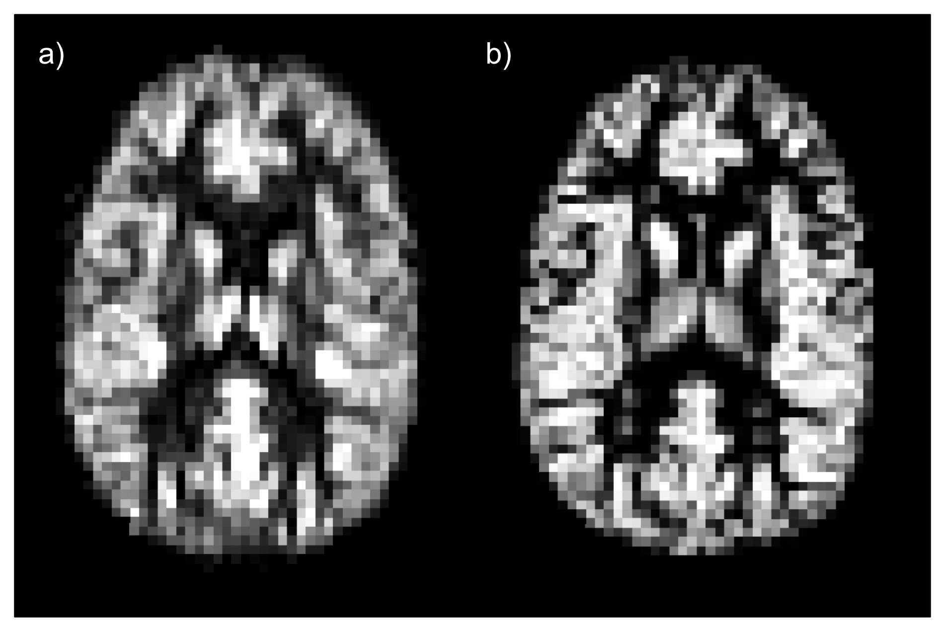
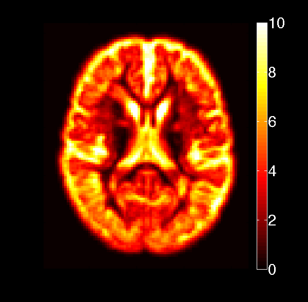
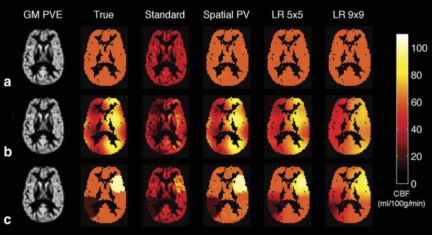
The mismatch in spatial resolution of Arterial Spin Labeling measurements of perfusion and the structure of functionally distinct tissues in the brain, such as grey and white matter, leads to biases in perfusion estimation known as Partial Volume Effects (PVE)1-3. PVE occur when the image voxels contain a mixture of tissue with distinct perfusion, as is common with typical ASL image resolution, leading to perfusion values that reflect the proportion of tissue in the voxel. It is already recognized that PVE influences studies of brain perfusion and its effect might be even more evident in studies where changes in perfusion are co-incident with variations in brain structure, such as studies involving a comparison between a patient population vs control subjects or study-subjects over a large range of ages. A number methods exist for correction of PVE (PVEc)2,3; however, their use is currently limited and the methodologies employed in different studies are often inconsistent. This educational e-poster firstly seeks to demonstrate the influence of PVE on individual perfusion images, as well as the influence it will have in studies involving perfusion, and methods that are currently available that seek to correct for PVE and how they can be used.
Outline of Content
Physiological interpretation of perfusion and PVE - what do we mean by perfusion and is it reasonable to separate the perfusion contribution from different types of tissue?
The influence of PVE on ASL perfusion images - demonstration on the profound way that brain structure influences the look of perfusion images, as well as how it significantly alters measured perfusion values at the individual and group level.
Correcting for partial volume effects - The underlying theoretical rationale for PVEc methods and the pros and cons of the methods currently available.
Confounds for PVEc - An overview of some of the major effects that can confound PVEc and where caution should be exercised.
Summary
PVE is an issue for ASL perfusion studies that will not go away until we achieve an order of magnitude improvement in resolution. Methods now exist for correction of PVE, but these are still not widely employed, meaning that our experience with them remains limited. Increasing availability of tools that implement these methods will allow more studies to use PVEc and thus let us understand whether wider adoption of such approached is required.Acknowledgements
We thank the European Union COST agency (through COST Action BM1103) for sponsorship of the workshops that have stimulated the discussion from which this e-poster has emerged.References
1. Alsop et al. Recommended implementation of arterial spin-labeled perfusion MRI for clinical applications: A consensus of the ISMRM perfusion study group and the European consortium for ASL in dementia. Magn Reson Med 73, 102–116, 2015.
2. Asllani et al. Regression algorithm correcting for partial volume effects in arterial spin labeling MRI. Magn Reson Med 60, 1362–1371, 2008.
3. Chappell et al. Partial volume correction of multiple inversion time arterial spin labeling MRI data. Magn Reson Med 65, 1173–1183, 2011.
Figures