4188
Brain Structural Network of Working Memory and Processing Speed for ALL Survivors1St Jude Children's Research Hospital, Memphis, TN, United States
Synopsis
Acute lymphoblastic leukemia survivors may have significant deficits in processing speed and working memory even when treated with only chemotherapy. We investigate the relationship of diffusion tensor imaging metrics in an a priori brain structural network with neurocognitive functions such as processing speed and working memory. We found that fractional anisotropy values in the structural network were significantly positively associated with processing speed performance in two MR exams two years apart, and axial diffusivity values were negatively associated with working memory in the MR exam at the end of therapy. These findings may provide potential evidence for a structural neurocognitive network.
Purpose
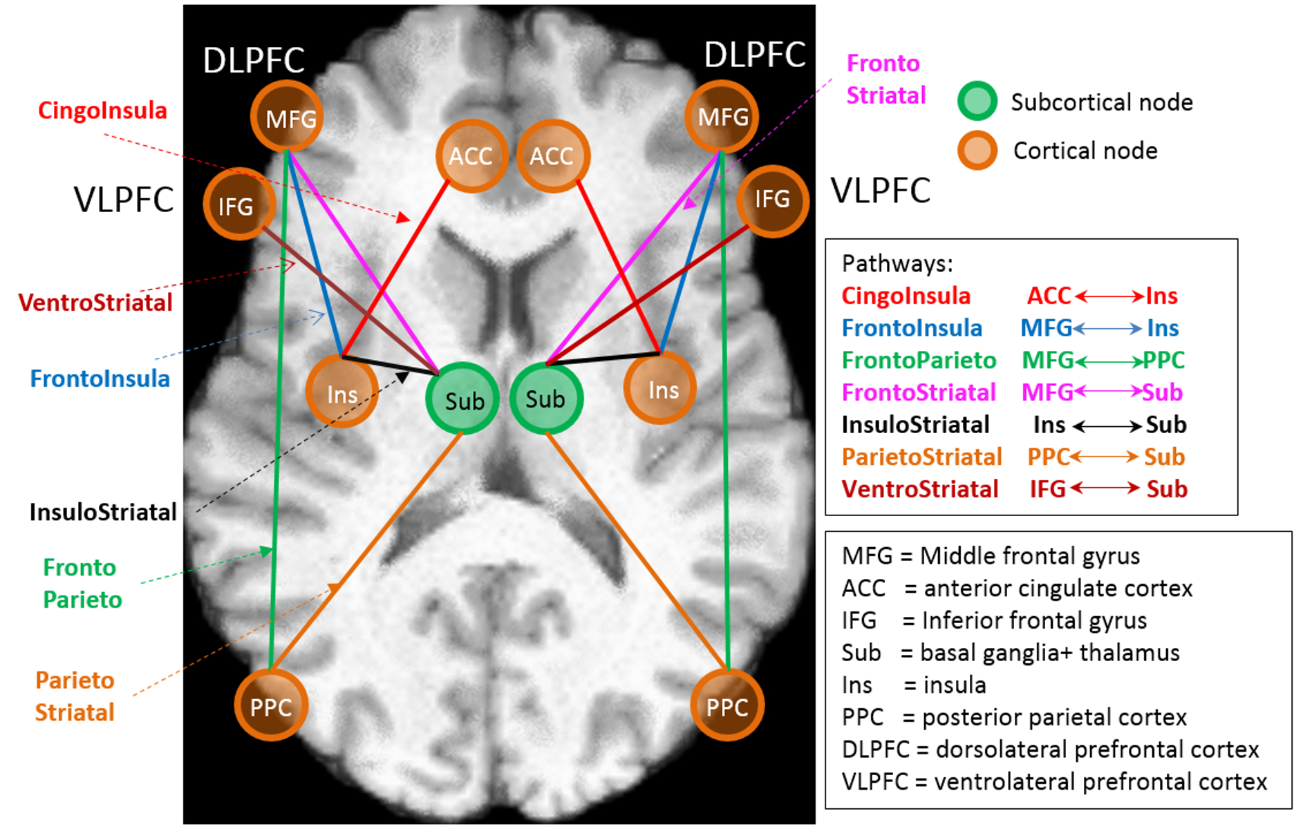
Acute lymphoblastic leukemia (ALL) is the most common form of cancer in childhood and adolescence. 5-year survival rates have improved to above 90% with contemporary chemotherapy alone. However, the chemotherapy drugs have also been associated with chronic neurotoxicity, which can result in impaired neurocognitive functions such as worse working memory and slower processing speed. Previously, we built a structural cognitive network from functional networks (central executive network, salience network, and subcortical network) based on fMRI studies (1-7). This structural cognitive network mainly includes dorsolateral prefrontal cortex (DLPFC), posterior parietal cortex (PPC), anterior cingulate cortex (ACC), insula, and subcortical cortex (basal ganglia and thalamus). To compensate for the low angular and spatial resolution of the clinically acquired diffusion tensor imaging (DTI) data, we created structural connectivity pathways using high resolution DTI data from the Human Connectome Project (HCP). In a previous preliminary study with a single MRI exam from 143 ALL survivors at end of chemotherapy, we found that DTI metrics in most pathways were significantly associated with working memory and processing speed performance. Here, we expanded our study to include 200 ALL survivors each with two MRI exams to further explore the association of DTI metrics with neurocognitive performance.Methods
Two MRI exams were acquired two years apart early in therapy at re-induction I and the end of therapy in 200 ALL survivors (121 male, 79 female, age at MR1 (re-induction) 7.2±4.4 , age at MR2 (end of therapy)/ Cognitive Assessment 9.4±4.4 years). All subjects were treated on a chemotherapy-only protocol, Total Therapy Study XVI (NCT00549848). Low resolution DTI data was acquired with 12 directions and a spatial resolution of 1.8 ×1.8 ×3.0 mm on a Siemens 3T scanner due to the time limitation and were processed with SPM8 (fil.ion.ucl.ac.uk/spm/).
We evaluated all subjects using a pre-built cognitive network (Fig. 1) using high-resolution DTI data from the Human Connectome Project (7). DTI metrics such as mean values of fractional anisotropy (FA), axial diffusivity (AD), and radial diffusivity (RD) were quantified in each pathway for 200 ALL survivors and 81 adult healthy volunteers from the HCP. Processing speed was assessed using the WJ-III Decision Speed test while working memory was assessed using the WJ Auditory Working Memory test and the Digit Span Backward (DSB) test.
A multiple linear regression model was used to fit the data. The model can be expressed as
$$Y_i=\beta_0+\beta_1DTI_i+\beta_2Age_i+\varepsilon_i$$
where $$$Y_i$$$ is a neurocognitive measure for the i-th subject. We included age as a covariate to adjust for the age effect on the neurocognitive measures as well as DTI measures. We applied the false discovery rate (FDR) correction procedure of Benjamini and Hochberg to correct the p-values for multiple testing. Results were considered significant at the P < 0.05 level.
Results
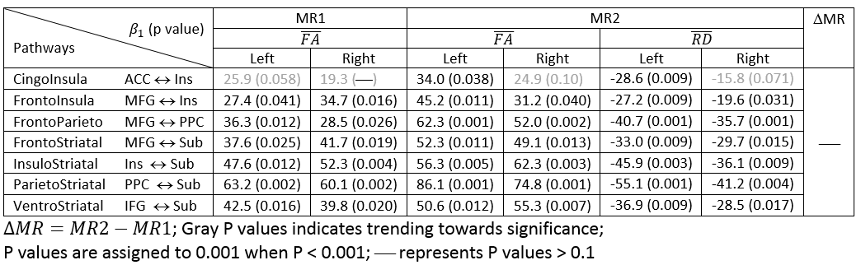
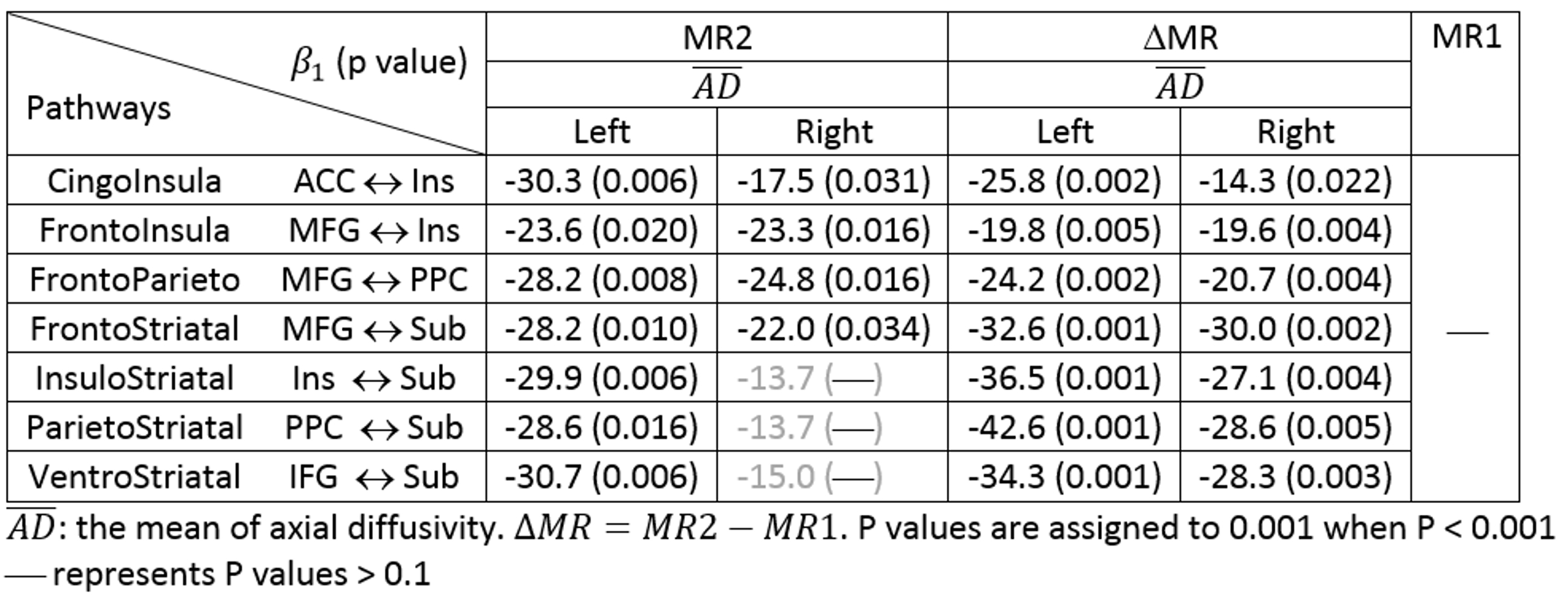
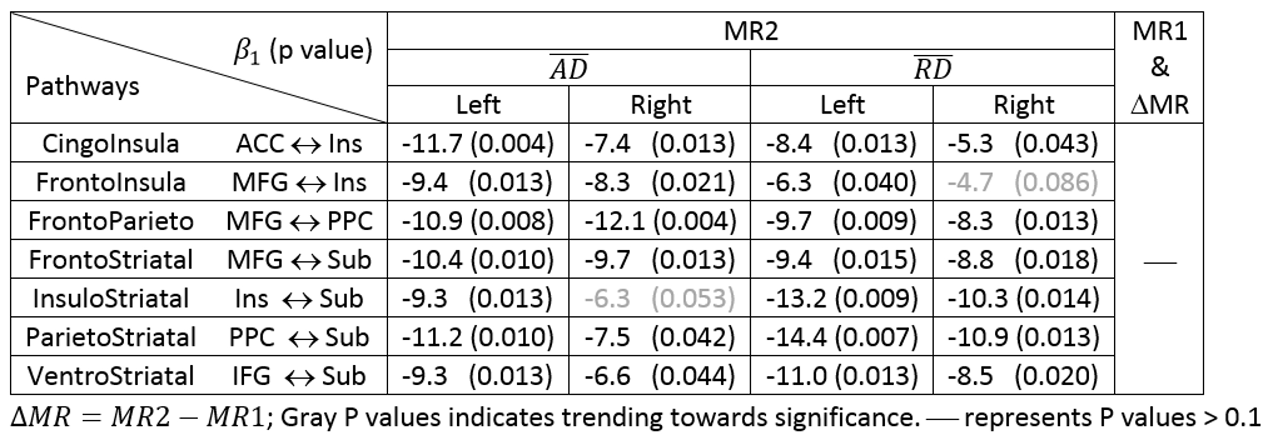
The mean FA values in both MR1 and MR2 were significantly associated with WJ-III Decision Speed in most of pathways bilaterally (Table1). The mean RD was significantly associated with Decision Speed only at MR2, but not at MR1. The mean AD was significantly associated with WJ Auditory Working Memory scores in most of pathways at both MR2 and ΔMR (Table 2). Consistently, the mean AD values in most of pathways at MR2 were significantly associated with DSB measures, which also assess working memory (Table 3). In addition, the mean RD at MR2 shows significant association with DSB measures. Neither mean AD nor RD showed any significant association with DSB measures at MR1 and ΔMR (Table 3). None of these DTI metrics had any significant association with the processing speed and working memory measures in adult volunteers.Discussion / Conclusion
Both larger FA and smaller RD may indicate a higher degree of myelination in white matter. The thicker myelin sheaths enable signals to transmit faster in axons. These facts are consistent with our findings: a positive association of FA and a negative association of RD with processing speed. The association between FA and processing speed is so strong that it is evident at MR1, which was two years earlier than the cognitive assessment. In contrast, AD and RD were negatively associated with working memory at MR2. Increases in AD and RD may indicate declines in working memory ability due to brain injury as a result of CNS-directed chemotherapy. Our findings were consistent with the previous results from a smaller cohort of subjects, and provide potential evidence for a structural neurocognitive network, which could be further used for evaluating survivors at greatest risk for reduced neurocognitive outcomes.Acknowledgements
This work was supported by Cancer Center Support Grant P30 CA-21765 from the National Cancer Institute at the National institutes of Health,Bethesda, MD, and by the American Lebanese Syrian Associated Charities (ALSAC), Memphis, TN.References
1. Bressler S, Memon V. Large-scale brain networks in cognition: emerging methods and principles. Trends in Cognitive Sciences 14 (2010), 277-290.
2. Memon V. Developmental pathways to functional brain networks: emerging principles. Trends in Cognitive Sciences 17 (2013), 627-640.
3. Seeley, W. et al. Dissociable intrinsic connectivity networks for salience processing and executive control. J. Neuosci. 27 (2007), 2349-2356.
4. Frank M. et al. Interactions between frontal cortex and basal ganglia in working memory: a computational model. Cogn Affect Behav Neurosci. 1 (2001), 137-160.
5. Collette F. et al. Exploration of the neural substrates of executive functioning by functional neuroimaging. Neuroscience. 139 (2006), 209-221.
6. O’Reilly R. et al. Making working memory work: a computational model of learning in the prefrontal cortex and basal ganglia. Neural Comput. 18 (2006), 283-328.
7. Guo J. et al. DTI Association with Working Memory and Speed in Cognitive Network Pathways. Proc of ISMRM, 24th, p3443, Singapore, 2016.
Figures