4179
Clinical Applications of Simultaneous Multi-slice (SMS) Imaging with High-angular-resolution Diffusion Imaging (HARDI) : a Comparative Study of Brain Tumor Pre-operative Evaluation with and without SMS1Radiology, Université Laval, Québec, QC, Canada, 2Radiology, CHU de Québec, Québec, QC, Canada, 3CHU de Québec, QC, Canada
Synopsis
Diffusion MRI can be helpful in pre-operative brain tumor evaluation to assess white matter tracts involved. It is however a time-consuming sequence, a problem that Simultaneous Multi-slice Imaging was designed to solve, although it has to demonstrate its quality non-inferiority. We compared in five patients tractographic data (using HARDI), acquired with and without SMS techniques, to evaluate the quality of imaging data as measured by the number of fibers between two regions of interest. We found that SMS-factor 2, cut time down by 37%, which is clinically relevant. We also found that SMS-2 is indeed non-inferior (alpha=0,03125) and can be used without quality compromise.
Introduction
Diffusion MRI can be used in a clinical setting to evaluate the white matter involvement of a brain tumor before neurosurgery is undertaken, which allows the radiologist and neurosurgeon to plan maximal tumor resection with minimal functional impairment. One of the limitations of such a study is the acquisition time, in addition to the various other MRI sequences already needed. Simultaneous Multi-slice (SMS) imaging is meant to reduce acquisition time by performing parallel imaging, but we have to ensure that there is no compromise on the quality of resulting data. We therefore designed this study to compare, in patients with brain tumor and considering the time spared, the image quality of SMS imaging versus non-SMS imaging, to assess its non-inferiority and quantify the time spared.
Methods
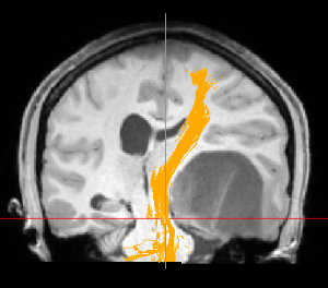
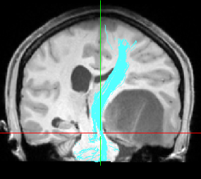
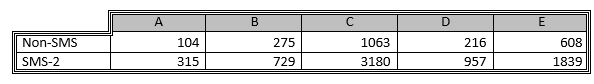
We recruited 5 patients who went through their pre-operative MRI and obtained Diffusion Imaging data, first without SMS and then with SMS factor 2, using a 3T MRI device, in our tertiary neurological institute. Time acquisition was measured for both non-SMS and SMS-2. We then used High-angular-resolution Diffusion Imaging (HARDI) to identify white matter tracts going from a proximal (to the tumor) region of interest (ROI) to a distal one. To evaluate the quality of data as a radiologist would judge it, we obtained the number of tracts identified in similar ROIs in each technique; indeed, more tracts are easier to interpret for the clinician as well as enhance the precision of the clinical pre-operative planning. Statistical analysis was performed with the five patients by using the binomial test to compare the non-SMS results to the SMS-2 results.Results
First, we found that, without SMS, acquisition time is 10min 41sec; with SMS-2 it is 6min 42sec. This represents a 3min 59sec reduction (or 37,29%), for this sequence alone.
Second, we found that for all five patients the number of fibers was actually superior in SMS-2 than in non-SMS Imaging. The binomial test shows that the alpha-value is of 3.125% for non-inferiority, under the 5% threshold of statistical significance.
Discussion
Compared to non-SMS technique, using Simultaneous Multi-slice Imaging has allowed to save 37% of acquisition time for the diffusion Imaging sequence, a clinically relevant amount of time, given the already long time needed to do a full pre-operative MRI session in patients with brain tumor. This time reduction would benefit the patient, but would also improve the device use efficiency.
Moreover, the binomial test confirms, as for the number of fibers in tracts going between two similiar ROIs using HARDI, the non-inferiority (alpha=0,03125) of the SMS-2 imaging technique compared to conventional non-SMS technique. It indicates that quality is not undermined by the parallel imaging process. Data actually shows that there might be a tendency to superiority, though this was not the point of the study.
Conclusion
In conclusion, SMS-2 imaging has shown a benefit on time acquisition for the Diffusion Imaging sequence and was demonstrated as being non-inferior to conventional non-SMS technique qualitatively. It is therefore clinically relevant to use this technique, for example in pre-operative evaluation of the white matter involvement of brain tumors. Other research should be undertaken on higher SMS factors (to evaluate if time reduction remains associated with non-inferior data quality); we should also investigate if SMS technique could inversely be used with the aim of enhancing quality, for a given acquisition time.Acknowledgements
No acknowledgement found.References
No reference found.Figures