4177
Diffusion kurtosis imaging: a novel tool to evaluate survival of glioma patients1Fujian Medical University Union Hospital, Fuzhou, People's Republic of China, 2Tongji Hospital, HUST, Wuhan, People's Republic of China
Synopsis
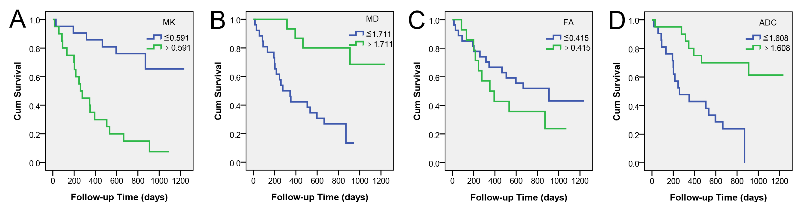
DKI is a promising tool to predict the survival of glioma patients. MK, MD and ADC were significantly correlated with overall survival (OS) of patients with astrocytic tumor. By univariate Kaplan-Meier survival analyses, OS of the patients was related to tumor grade, Ki-67 LI, resection status, enhancement degree, edge, edema degree, lesion number, MK, MD and ADC (log rank p < 0.05 for all). Multivariate Cox regression analysis indicated that MK is an independent predictor of OS in these patients, and it is a risk factor (P = 0.006, HR=2.142 and 95%CI=1.247-3.679 for MK increasing every 0.1). These results are helpful to clinic.
Objective
Survival time is important for glioma patients. Diffusion kurtosis imaging, an advanced non-Gaussian diffusion imaging technique, has shown great potential in grading gliomas and in assessment of proliferation of glioma cells. However, its role in evaluating survival of glioma patients has not been described. Therefore, the aim of this study is to evaluate the prognostic value of diffusion kurtosis imaging (DKI) for survival in patients with gliomas.Methods
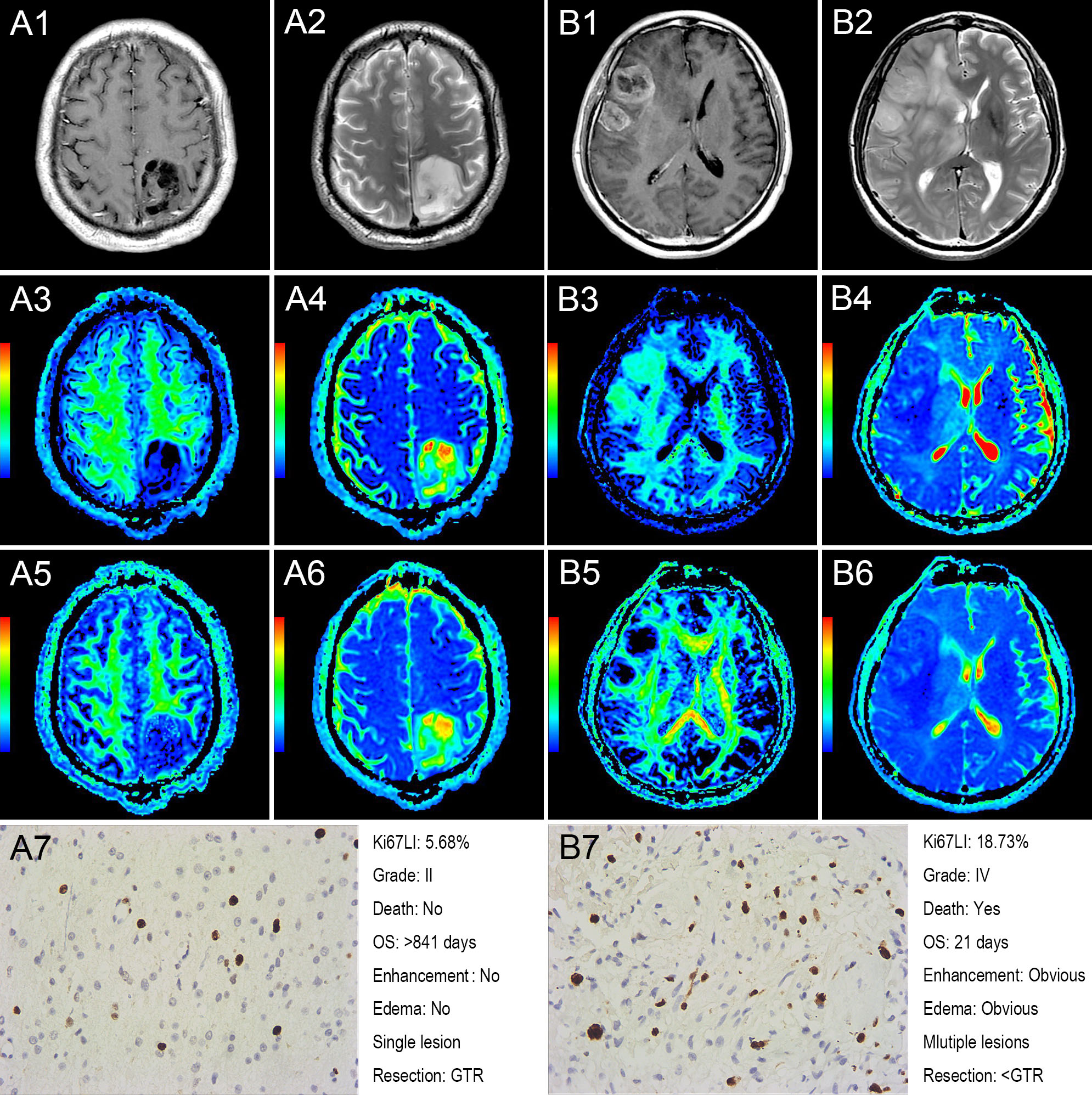
DKI, diffusion weighted imaging (DWI) and routine magnetic resonance imaging (MRI) were performed on 41 patients with confirmed primary astrocytic tumor. Clinical and pathological information including gender, age, KPS, tumor grade, Ki-67 LI, resection and chemoradiotherapy were recorded. Tumor characteristics including size, intensity, enhancement, edge, edema and lesion number also were evaluated from routine MRI. Mean kurtosis (MK), mean diffusivity (MD), fractional anisotropy (FA) and apparent diffusion coefficient (ADC) were subsequently calculated. Correlation analysis between overall survival (OS) and diffusion metrics was first performed for 24 dead patients. Univariate analysis (Kaplan-Meier survival analysis) between the OS and each factor were further performed, followed by a multivariate Cox regression analysis including gender, age, Karnofsky performance status (KPS), resection status, MK and ADC.Results
MK, MD and ADC were significantly correlated with OS of patients with astrocytic tumor (P<0.05). By univariate Kaplan-Meier survival analyses, OS of the patients was related to tumor grade, Ki-67 LI, resection status, enhancement degree, edge, edema degree, lesion number, MK, MD and ADC (log rank p < 0.05 for all). Multivariate Cox regression analysis indicated that MK is an independent predictor of OS in these patients, and it is a risk factor (P = 0.006, HR=2.142 and 95%CI=1.247-3.679 for MK increasing every 0.1).Conclusion
DKI is a promising tool to predict the survival of glioma patients, and MK is an independent predictor of overall survival, which is helpful to clinic.Key words
Diffusion Kurtosis Imaging; Glioma; Survival AnalysisAcknowledgements
The authors thank Wenzhen Zhu for her advice on manuscript writing, thank Hao Zhang, Haijun Sang and Siquan Wang for their assistance with the follow-up of the patiens, and also thank Chanchan Liu for his assistance with the statistical analyses.References
1. Federau, C., et al., IVIM perfusion fraction is prognostic for survival in brain glioma. Clin Neuroradiol, 2016.
2. Saksena, S., et al., Predicting Survival in Glioblastomas Using Diffusion Tensor Imaging Metrics. Journal of Magnetic Resonance Imaging, 2010. 32(4): p. 788-795.
3. Jiang, R., et al., Diffusion kurtosis imaging can efficiently assess the glioma grade and cellular proliferation. Oncotarget, 2015. 6(39): p. 42380-93.
4. Louis, D.N., et al., The 2007 WHO classification of tumours of the central nervous system. Acta Neuropathol, 2007. 114(2): p. 97-109.
Figures
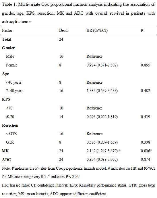
Table 1: Multivariate Cox proportional hazards analysis indicating the association of gender, age, KPS, resection, MK and ADC with overall survival in patients with astrocytic tumor.
Note: P indicates the P value from Cox proportional hazards model. # indicates the HR and 95%CI for MK increasing every 0.1. * indicates P < 0.05. HR: hazard ratio; CI: confidence interval; KPS: Karnofsky performance status, GTR: gross total resection; MK: mean kurtosis; ADC: apparent diffusion coefficient.