4169
Evaluation of the Applicability of Territorial Arterial Spin Labelling in Meningiomas for Presurgical Assessments Compared with 3-Dimensional Time-of-Flight Magnetic Resonance Angiography1Department of Radiology, Huashan Hospital, Fudan University, Shanghai, People's Republic of China, 2Department of Neurosurgery, Huashan Hospital, Fudan University, Shanghai, People's Republic of China, 3Department of Radiology, Shanghai Cancer Center, Shanghai, People's Republic of China, 4MR Research China, GE Healthcare, Shanghai, People's Republic of China
Synopsis
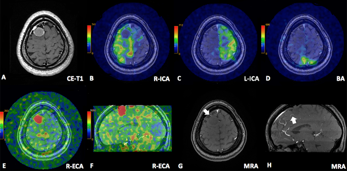
In this research, territorial Arterial Spin Labelling (t-ASL) and unenhanced 3D-TOF-MRA were used to evaluate the usage in identification of the feeding vasculature of meningiomas in 20 consecutive patients. Results showed that the inter-observer agreement was excellent for the identification of the origin of the feeding arteries by t-ASL, which was better than the inter-observer agreement of 3D-TOF-MRA. The inter-modality agreement between t-ASL and 3D-TOF-MRA for the feeding arteries was moderate. The information about feeding arteries was potentially related to patients’ symptoms and pathology, making it crucial for neurosurgeons in planning surgery and evaluating prognosis.
Purpose
Meningiomas are generally known as hyper-perfusion tumours mainly supplied by branches of the extra-carotid artery (ECA) and intra-carotid artery (ICA). The improper clipping or embolization may cause permanent ischemic injury to the brain cortex or nerves, resulting in blindness, dyskinesia, sensory disturbances, etc. Thus a comprehensive information of the feeding arteries of meningiomas is helpful for optimizing treatment strategy and avoiding severe complications. DSA, as the gold standard, is limited in usage as of high expense, surgical risk, and invasiveness. MR Angiography, especially Time of Flight MRA (TOF) has emerged as a replacement to depict the arterial architecture [1-2]. However, it remains difficult to distinguish the origin of small vessels around the tumour as lack of vessel selectivity. Territorial arterial spin labeling (t-ASL), capable of linking feeding arteries and perfusion region, has been introduced. This study aims to prospectively evaluate the application of t-ASL in comparison with unenhanced 3-dimensional Time-of-Flight Magnetic Resonance Angiography (3D-TOF-MRA) in the identification of the feeding vasculature of meningiomas.Methods
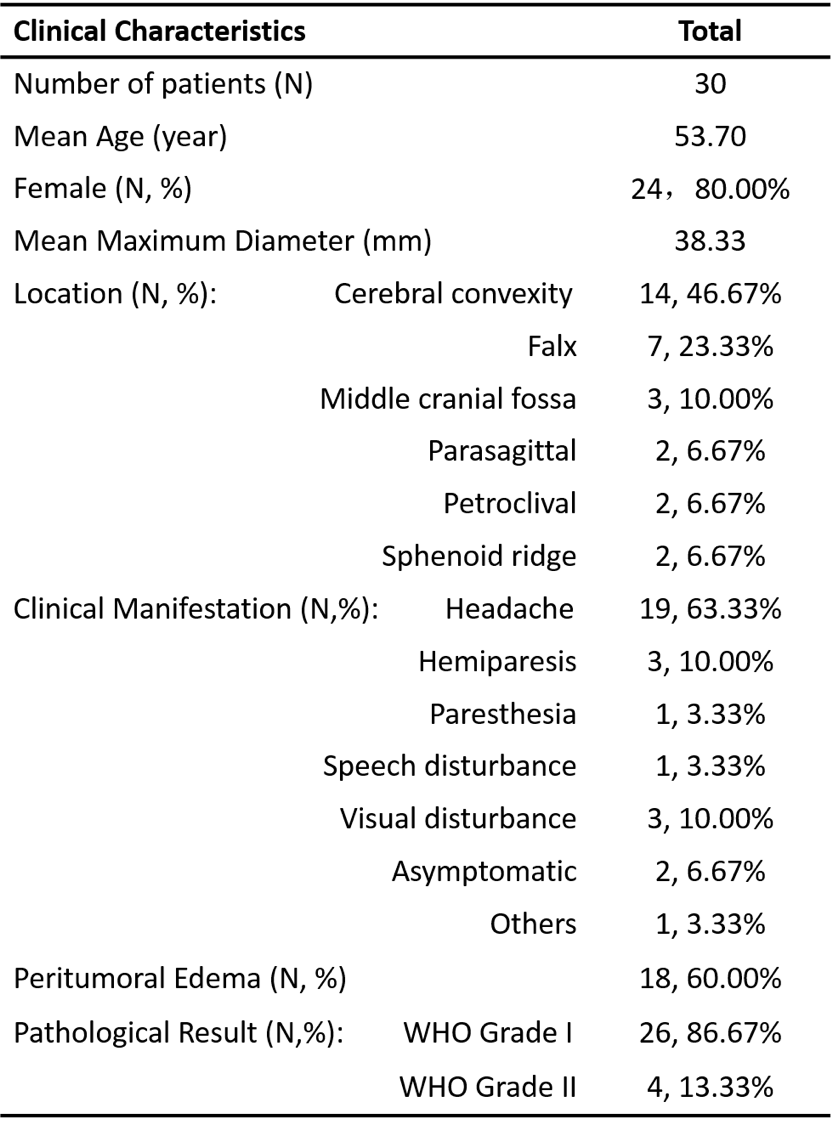
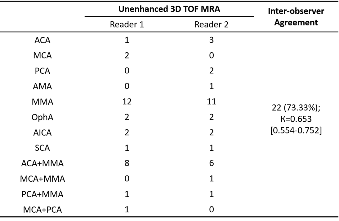
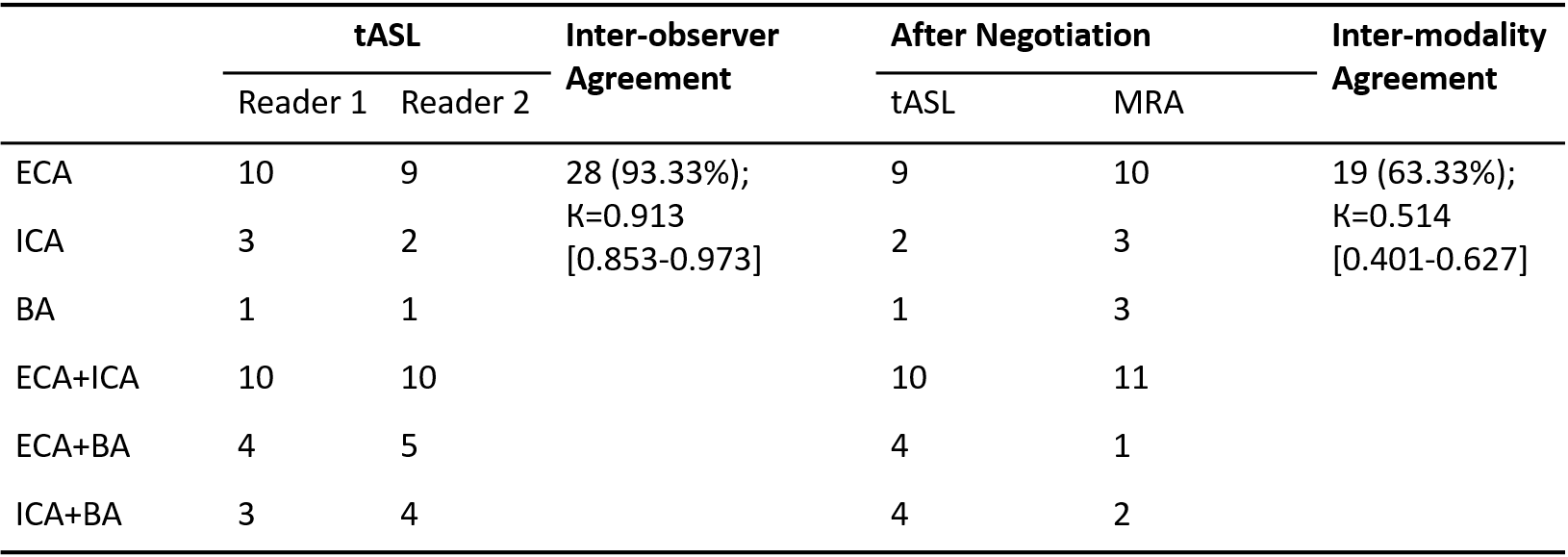
Thirty consecutive patients with suspected meningiomas underwent conventional MR imaging, unenhanced 3D-TOF-MRA and t-ASL perfusion imaging on a 3.0 T scanner (GE, DISCOVERY MR750W). TOF-MRA was carried out with the following parameters: TR/TE/NEX 8.2 ms/3.2 ms/1, flip angle 12°, and effective voxel size, 0.75×0.75×1.0 mm3. The t-ASL were acquired using a 3D FSE with stack of spiral readout with the following parameters: arms/points per arm 4/512, FOV 20×20 cm2, slice thickness 4 mm, labelling duration 1.45s, and post-labelling delay 1.5s. Super-selective scheme was used for vessel selective labeling. The feeding vessels with each technique were evaluated by 2 experienced neuro-radiologists. Cohen’s к statistics was applied to evaluate the diagnostic performance of both techniques. The levels of inter-observer agreement and inter-modality agreement (between consensus readings of MRA and t-ASL images) with regard to the feeding arteries were determined by the following criteria: к<0.20, poor; к=0.21–0.40, fair; к= 0.41–0.60, moderate; к= 0.61–0.80, good; к= 0.81–0.90, very good; and к> 0.90, excellent agreement.Results
For the identification of the origin of the feeding arteries on t-ASL, the inter-observer agreement was excellent (к=0.913), while the inter-observer agreement of 3D-TOF-MRA was good (к=0.653). The inter-modality agreement between t-ASL and 3D-TOF-MRA for the feeding arteries was moderate (к=0.514). All 8 patients with motor or sensory disorders proved to have meningiomas supplied completely or partially by the internal carotid arteries, while all 14 patients with meningiomas supplied by the external carotid arteries or basilar arteries didn`t show any symptoms concerning motor or sensory disorders (p=0.003).Discussion
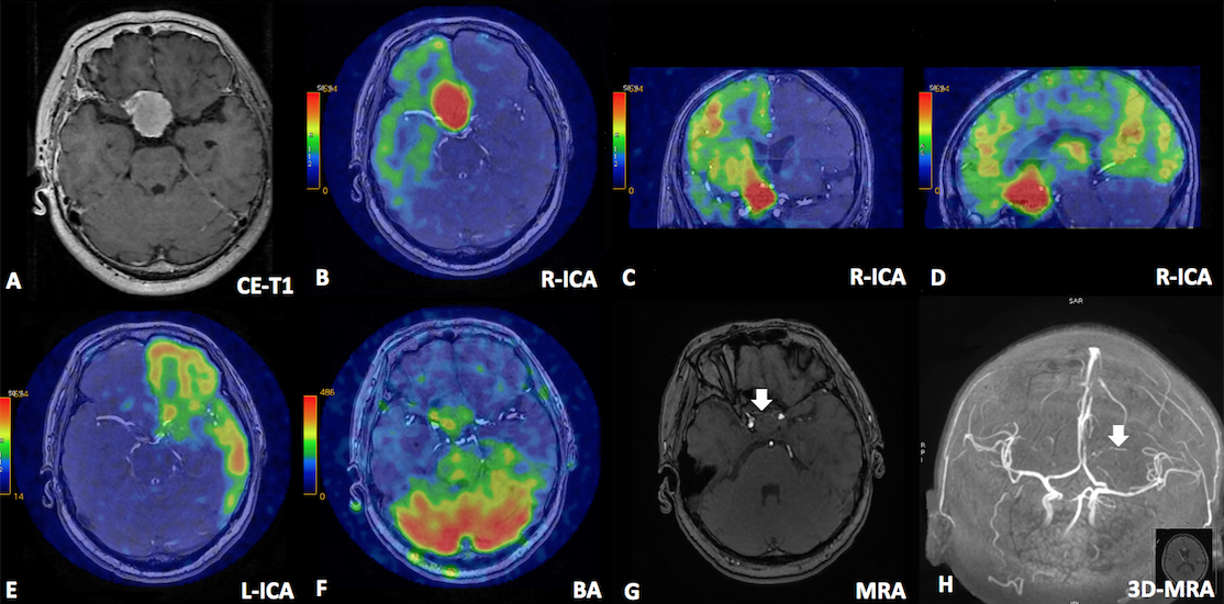
Knowledge of the exact feeding arteries is of great importance in the understanding of and operations for meningiomas. In our research, MRA could provide information about the morphology of surrounding arteries but with no sufficient proof of which blood supply artery was. Territorial ASL, on the contrary, achieved excellent inter-agreement when judging the origin of the artery branches, indicating that it was relatively easy for radiologists to distinguish mono-blood-supplied tumors from double- or even multiple blood vessel-supplied tumors by territorial ASL. The clinical manifestation can indicate the blood supply of meningioma. According to our results, all of the meningiomas with manifestations of motor or sensory disorders were fed totally or partially by the ICA, with sensitivity of 50.00%,specificity of 100%, PPV of 100%, and NPV of 63.64%. Furthermore, all four of the atypical meningiomas in our study were supplied by double arteries, and the ICA was the main feeding artery in all of these cases, which indicated that feeding arteries might be related to the pathology and prognosis of the tumor; further research is encouraged.Conclusion
T-ASL could precisely evaluate the origin of arterial supply of meningioma, in a safe, intuitive, non-radioactive manner. Since its limitation to label small vessels, t-ASL could serve as a complement to unenhanced 3D-TOF-MRA and increase accuracy in the identification of the supplying arteries of meningiomas. The information about feeding arteries was potentially related to patients’ symptoms and pathology, making it more crucial for neurosurgeons in planning surgery as well as evaluating prognosis.Acknowledgements
No acknowledgement found.References
[1] Iryo Y, Hirai T, Kai Y, Nakamura M, Shigematsu Y, Kitajima M, et al. Intracranial dural arteriovenous fistulas: evaluation with 3-T four-dimensional MR angiography using arterial spin labeling. Radiology 2014;271(1):193-9.
[2] Uetani H, Akter M, Hirai T, Shigematsu Y, Kitajima M, Kai Y, et al. Can 3T MR Angiography Replace DSA for the Identification of Arteries Feeding Intracranial Meningiomas? American Journal of Neuroradiology 2013;34(4):765-772.
Figures