4164
The association of MRI-detected brain volume change with prenatal exposure to Perfluoroalkyl Substances in teenage1Institute of Medicine, Chung Shan Medical University, Taichung, Taiwan, 2Department of Medical Imaging and Radiological Sciences, Chung Shan Medical University, Taichung, Taiwan, 3Department of Medical Imaging, Chung Shan Medical University Hospital, Taichung, Taiwan, 4National Institute of Environmental Health Sciences, National Health Research Institutes, Miaoli, Taiwan, 5School of Medicine and Department of Pediatrics, Chung Shan Medical University and Hospital, Taichung, Taiwan
Synopsis
The current study was to explore the relationship between prenatal exposure to PFASs, determined in maternal blood collected during the third trimester of pregnancy and the children’s brain volume difference at the age of 13-15 years old. The results showed a significant negative correlation between the maternal blood PFASs concentrations and the children's brain MRI in multiple different brain areas, including both gray matter and white matter.
INTRODUCTION
Perfluoroalkyl Substances (PFASs) have been produced more than a half-century and widely used in modern commercial and industrial products as an aid in processing polymers, surface coatings, pharmaceuticals, and surfactants in cleaning products. PFASs are highly resistant to chemical, thermal, biological degradation and can migrate from the products and enter the environment1. Animal and human exposures are majorly through polluted food/water and air/dust intake. Exposure to PFASs, especially during early life, could be harmful and result in a range of adverse health effects, including disturbed immune and neuroendocrine system, also hepatic and neuro-toxicity2. Multiple neurobehavioral effects have been observed in animal and human studies, but the evidence of brain image associated with PFASs effect in human is lacking. In this study, we investigated the associations between prenatal exposure to PFASs and MRI brain volume difference in teenage.METHODS
We recruited 47 mother–child pairs from the general population (25male, 22 female) in central Taiwan for this study. We examined the association between PFASs in the maternal blood collected during the third trimester of pregnancy and the children's brain MRI at 13 to 15 years of age (mean=13.9). The examined PFASs were including perfluorooctane sulfonate (PFOS), perfluorooctanoic acid (PFOA), perfluorononanoic acid (PFNA), perfluoroundecanoic acid (PFUA) and perfluorododecanoic acid (PFDoA). All images were acquired using a 3-Tesla MRI (Skyra, Siemens, Germany) with a 20 channel head-neck coil. All structural images were acquired using the three-dimension magnetization-prepared rapid gradient-echo (3D MPRAGE) sequence with TR/TE/TI=2500/2.27/902 ms, flip angle=8°, resolution=0.98×0.98×1 mm2, and total slices=160. In Voxel-based morphometry (VBM) analysis, all structural data were processed using Statistical Parametric Mapping 8 (SPM8, Wellcome Department of Cognitive Neurology, London, UK) with Voxel-Based Morphometry 8 (VBM8, University of Jena, Department of Psychiatry, Jena, Germany) toolbox. Noise reduction was performed first using spatial adaptive non-local means. Subsequently, whole-brain native space T1WI were segmented into gray matter, white matter, and cerebrospinal fluid components, and then imported into a rigidly aligned space and iteratively registered to the East Asian brain model (implemented in VBM8). Associations between the maternal blood PFASs concentrations and the teenage brain MRI were estimated using multiple regression analysis by SPM8 with adjustments for the teenage sex and whole brain volume. Standard T1WI were implemented in SPM8 as the underlying map. A P-value less than 0.05 was considered statistically significant.RESULTS
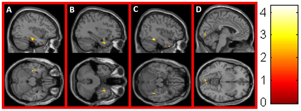
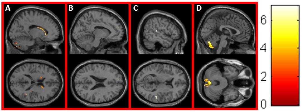
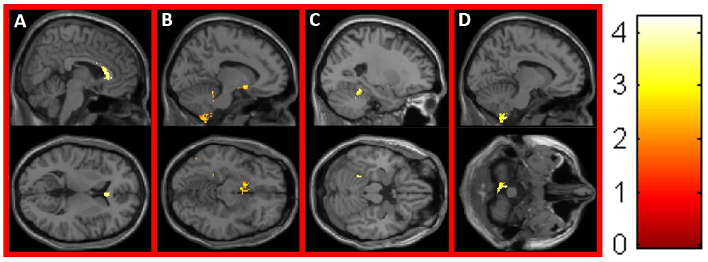
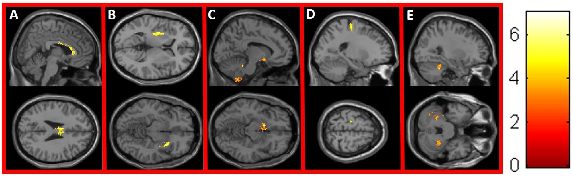
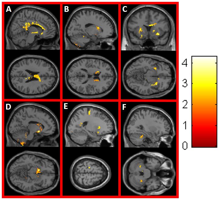
The mean PFASs concentrations in the maternal blood were PFOS=14.85 ng/mL, PFOA=2.93 ng/mL, PFNA=1.70 ng/mL, PFUA=4.76 ng/mL, PFDoA=0.33 ng/mL. There were no significant differences in maternal blood PFASs concentrations, age or body mass index (BMI) between male and female. In association analysis between the maternal blood PFASs concentrations and brain MRI VBM, significant negative correlations (P<0.05) were noted between corpus callosum and PFNA/ PFUA/PFDoA (Fig 3A, 4A, and 5A); between external capsule and PFUA/PFDoA (Fig 4B and 5C); between caudate nucleus and PFOA/PFDoA (Fig 2A and 5B); between hypothalamus and PFNA/ PFUA/PFDoA (Fig 3B, 4C, and 5D); between cerebellum and PFOA/PFNA/PFUA/PFDoA (Fig 2D, 3C, 3D, 4E, and 5F); between precentral gyrus and PFUA/PFDoA (Fig 4D and 5E); between Inferior temporal lobe/ occipital lobe and PFOS (Fig 1A-D); between frontal lobe/ temporal lobe and PFOA (Fig 2B, 2C).DISCUSSION
The current study was to explore the relationship between prenatal exposure to PFASs, determined in maternal blood collected during the third trimester of pregnancy and the children’s brain volume difference at the age of 13-15 years old. The results showed a significant negative correlation between maternal blood PFASs concentrations and the children's brain MRI in multiple different brain areas. The findings may support the previous studies that PFASs are able to cross the blood–brain barrier (BBB) and can cause disruption to the central nervous system3-5. The most well-known PFASs are perfluorosulfonates (PFSAs) and perfluorocarboxylates (PFCAs). We found that PFCAs (PFOA, PFNA, PFUA and PFDoA) are negative correlation majorly in deep regions of the brain, in contrast, PFSAs (PFOS) are negative correlation in inferior temporal and occipital lobes. The findings may indicate that PFCAs can cross the BBB more efficiently than PFSAs and affect deep brain structure3. Furthermore, we found a significant negative correlation between PFASs concentration and precentral gyrus and cerebellum, which may support the previous studies that prenatal exposure of PFSAs can affect motor function and even increased the risk of congenital cerebral palsy6,7.CONCLUSION
In this teenage brain MRI VBM study, we revealed that prenatal exposure of PFASs may cause multifocal brain volume decrease. We suggested that PFCAs seemed to affect deep brain structure more than PFSAs and increased prenatal PFASs exposure result in precentral gyrus and cerebellum volume decreased, which may affect motor function.Acknowledgements
This study was supported in part by the nationwide Taiwan Maternal and Infant Cohort Study.References
1. Houde M, Martin JW, Letcher RJ, et al. Biological monitoring of polyfluoroalkylsubstances: a review. Environ Sci Technol. 2006; 40(11): 3463–3473.
2. Yu N, Wei S, Li M, et al. Effects of Perfluorooctanoic Acid on Metabolic Profiles in Brain and Liver of Mouse Revealed by a High-throughput Targeted Metabolomics Approach. Sci Rep. 2016; 1; 6: 23963.
3. Greaves AK, Letcher RJ, Sonne C, et al. Brain region distribution and patterns of bioaccumulative perfluoroalkyl carboxylates and sulfonates in east greenland polar bears (Ursus maritimus). Environ Toxicol Chem. 2013; 32(3): 713-22.
4. Wang FQ, Liu W, Jin YH, et al. Transcriptional effects of prenatal and neonatal exposure to PFOS in developing rat brain. Environ Sci Technol. 2010; 1; 44(5): 847-53.
5. Johansson N, Fredriksson A, Eriksson P. Neonatal exposure toperfluorooctane sulfonate (PFOS) and perfluorooctanoic acid (PFOA)causes neurobehavioural defects in adult mice. Neurotoxicology. 2008; 29(1): 160-9.
6. Chen MH, Ha EH, Liao HF, et al. Perfluorinated compound levels in cord blood and neurodevelopment at 2 years of age. Epidemiology. 2013; 24(6): 00-8.
7. Liew Z, Ritz B, Bonefeld-Jørgensen EC, et al. Prenatal exposure to perfluoroalkyl substances and the risk of congenital cerebral palsy in children. J.Am J Epidemiol. 2014; 15; 180(6): 574-81.
Figures