4163
Assessment of chemotherapy-induced brain structural alterations in breast cancer patients using GQI and its correlation with cognitive performance1Department of Medical Imaging and Radiological Sciences, Chung Shan Medical University, Taichung, Taiwan, 2Department of Radiology, Taichung Veterans General Hospital, Taichung, Taiwan, 3School of Medicine, Chang Gung University, Taoyuan, Taiwan, 4Department of Psychiatry, Chang Gung Memorial Hospital, Chiayi, Taiwan, 5Department of Psychology, Chung Shan Medical University, Taichung, Taiwan, 6Breast Center, Taichung Tzu Chi Hospital, Taichung, Taiwan, 7Department of Medical Imaging, Chung Shan Medical University Hospital, Taichung, Taiwan
Synopsis
Neuroimaging studies suggest that white matter and cognitive function changes were affected by breast cancer and its treatments. Our study interested in the early effect of the brain by chemotherapy. This study included 19 breast cancer survivors who had completed their chemotherapy and 20 healthy control group. Generalized q-sampling imaging (GQI) with voxel-based analysis was performed to show the brain structural differences between two groups. Multiple regression was also used to detect the correlation between Mini-Mental State Examination (MMSE) and the indices of GQI. Our results provided further evidence that breast cancer and adjuvant chemotherapy are associated with adverse effects on white matter.
INTRODUCTION
Breast neoplasms cases are the most common cancer of women in Taiwan. Nowadays the Advances in cancer treatments have resulted in significantly improved survival rates, but are often associated with side effects, such as cognitive decline, that may reduce the quality of life. Neuroimaging studies suggest that white matter and cognitive function changes were affected by breast cancer and its treatments1. However, the previous studies focused on the late effect of the brain by chemotherapy. Our study interested in the early effect of the brain by chemotherapy. Since diffusion tensor imaging (DTI) is associated with restrictions in the resolution of crossing fibers, we tried to use generalized q-sampling imaging (GQI) that can overcome these difficulties and is advantageous over DTI for the tractography of the fiber bundle2.METHODS
This study included 19 women with a history of breast cancer who had completed their primary chemotherapy less than 6 months before the study entry and were currently without evidence of active cancer. The chemotherapeutic drugs they used are Taxotere and Epirubicin only. We have another 20 healthy women as control group. All participants underwent MRI on a 1.5T scanner (Aera, Siemens, Germany). For diffusion imaging, we performed a single shot, diffusion-weighted spin echo-planar imaging sequence with parameters: repetition time (TR) = 7200 msec, echo time (TE) = 107 msec, resolution = 2 x 2 x 4 mm3, b-values = 0, 1000, 2000 sec/mm2 in 129 noncollinear directions, number of excitations (NEX) = 1, and acquisition time was 16 min. All participants were evaluated by the Mini-Mental State Examination (MMSE), which tests five cognitive functions, including orientation, registration, attention and calculation, recall, and language. Diffusion imaging was first corrected for eddy current by FSL. Probability distribution function was reconstructed using GQI with DSI studio. Independent t-test was performed with SPM to find the differences between two groups. Because a significant difference in age between two groups (p < 0.001) was found, we used age as covariates of no interest. Finally, multiple regression was used to detect the correlation between MMSE and the indices of GQI for all participants. GQI is a q-space reconstruction method that can reconstruct orientation distribution functions (ODF) from a variety of diffusion datasets. The GQI indices included generalized fractional anisotropy (GFA) and normalized quantitative anisotropy (NQA). GFA is defined as the standard deviation divided by the root mean square of the ODF, indicating a measurement of the anisotropy. NQA is defined as the normalized amount of anisotropic spins that diffuse along the fiber orientation3. The MMSE is a brief, quantitative measure of cognitive status in adults. It can be used to estimate the severity of cognitive impairment at a given point in time, and to document an individual's response to treatment4. The higher the MMSE score, the better cognitive performance.RESULTS
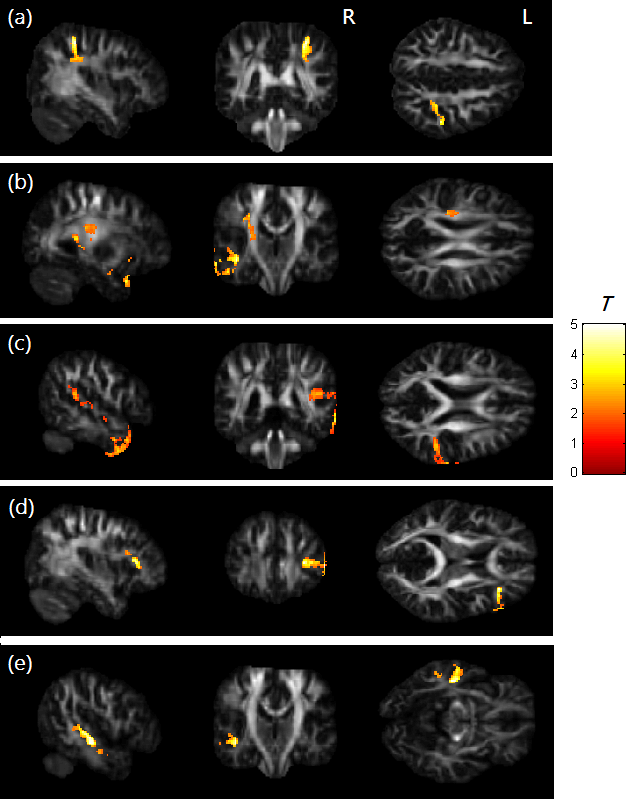
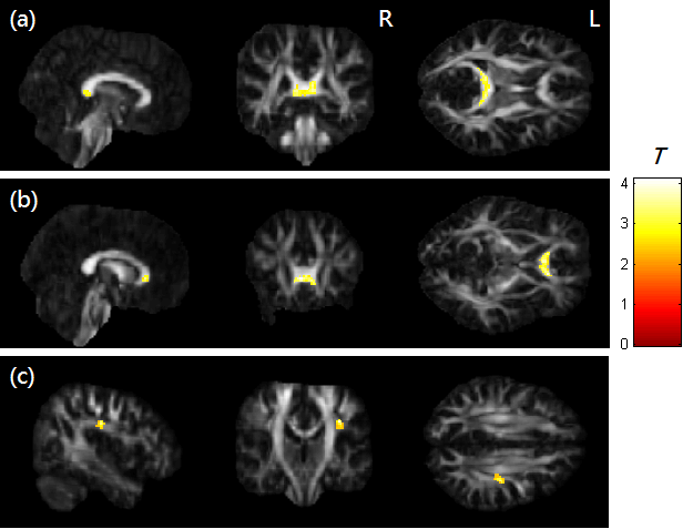
Compared to the control group, results from voxel-based analysis showed significantly lower GFA and NQA in the breast cancer group (p < 0.05). Differences of brain regions included right post-central blade, right superior temporal gyrus, right inferior frontal blade, left superior corona radiata and left middle temporal gyrus (Fig. 1). In addition, significant positive correlations between MMSE and GQI indices (GFA and NQA) were found in the regions of right superior corona radiata, splenium and genu of corpus callosum (p < 0.01) (Fig. 2).DISCUSSION
There are several potential reasons for white matter vulnerability and cognitive function decline after chemotherapy including toxicity of treatments to white matter5. White matter mediates communication among different brain regions and its integrity is important for optimal brain function. Damage to any part of the white matter connections can lead to changes in cognitive performance6. How Taxotere and Epirubicin cross the blood-brain barrier (BBB) remains a matter of debate. There is evidence that a frequently used chemotherapeutic agent, fluorouracil (FU), crosses the BBB by simple diffusion7,8. Systemic treatment with clinically relevant concentrations of FU in mice has been shown to cause damage to myelinated white matter tracts of the central nervous system (CNS)9.CONCLUSION
This study showed the decline of cerebral white matter and cognitive performance beyond chemotherapy. Our results provided further evidence that breast cancer and adjuvant chemotherapy are associated with adverse effects on white matter. Further research of this topic required larger samples and nonchemotherapy-exposed patients to determine the impact of brain organization.Acknowledgements
This study was supported in part by the research programs NSC103-2420-H-040-002 and MOST104-2314-B-040-001, which were sponsored by the Ministry of Science and Technology, Taipei, Taiwan.References
1. Hampson JP, Zick SM, Khabir T, et al. Altered resting brain connectivity in persistent cancer related fatigue. NeuroImage Clin. 2015; 8: 305–313.
2. Zhang H, Wang Y, Lu T, et al. Differences between generalized q-sampling imaging and diffusion tensor imaging in the preoperative visualization of the nerve fiber tracts within peritumoral edema in brain. Neurosurgery. 2013; 73: 1044–1053.
3. Yeh FC, Wedeen VJ, Tseng WY. Generalized q-sampling imaging. IEEE Trans Med Imaging 2010; 29: 1626–35.
4. Folstein MF, Folstein SE, McHugh PR. “Mini-Mental State”: a practical method for grading the cognitive state of patients for the clinician. J Psychiatr Res. 1975; 12: 189-198.
5. Dietrich, J. Chemotherapy associated central nervous system damage. Adv. Exp. Med. Biol. 2010; 678: 77e85.
6. Deprez S, Amant F, Smeets A, et al. Longitudinal assessment of chemotherapy-induced structural changes in cerebral white matter and its correlation with impaired cognitive functioning. J Clin Oncol. 2012; 30: 274-281.
7. Bourke RS, West CR, Chheda G, et al. Kinetics of entry and distribution of 5-fluorouracil in cerebrospinal fluid and brain following intravenous injection in a primate. Cancer Res. 1973; 33: 1735-1746.
8. Kerr IG, Zimm S, Collins JM, et al. Effect of intravenous dose and schedule on cerebrospinal fluid pharmacokinetics of 5-fluorouracil in the monkey. Cancer Res. 1984; 44: 4929-4932.
9. Han R, Yang YM, Dietrich J, et al. Systemic 5-fluorouracil treatment causes a syndrome of delayed myelin destruction in the central nervous system. J Biol. 2008; 7: 12.
Figures