4162
Susceptibility-related phase contrast associated with the alterations of myelo-architecture in adult-onset leukoencephalopathy with neuroaxonal spheroids and pigmented glia1Department of Biomedical Engineering, Ulsan National Institute of Science and Technology, Ulsan, Korea, Republic of, 2Department of Neurology, Pusan National University Yangsan Hospital, Yangsan, Korea, Republic of, 3Department of Electrical and Computer Engineering, Ulsan National Institute of Science and Technology, Ulsan, Korea, Republic of, 4Department of Forensic Medicine, Pusan National University Yangsan Hospital, Yangsan, Korea, Republic of, 5Department of Neurology, Pusan National University Hospital, Busan, Korea, Republic of, 6Department of Neurology, Pusan National University Yangsan Hospital
Synopsis
To investigate the origin of susceptibility-weighted imaging (SWI) contrast in frontal white matters of adult-onset leukoencephalopathy with neuroaxonal spheroids and pigmented glia (ALSP), we performed a combined post-mortem magnetic resonance imaging (MRI) and histological study of ALSP pathology. The myelin architectural changes, marked central myelin loss with preserved U-fibers beneath cortical gray matter, mainly contributed to the susceptibility contrast.
Purpose
Susceptibility-weighted gradient echo MRI techniques are sensitive in identifying white matter pathology in patients with multiple sclerosis (MS).1 Previous studies have shown iron depositions within activated microglia/macrophages in white matter MS lesions. Adult-onset leukoencephalopathy with neuroaxonal spheroids and pigmented glia (ALSP) is a hereditary leukodystrophy due to mutations in the colony stimulating factor 1 receptor (CSF1R) gene.2 ALSP has recently been considered one of the primary microglial disorders, characterized by non-inflammatory myelin loss, reactive astrocytosis, axonal spheroids, and pigmented microglia in the white matter. Subcortical U-fibers were generally spared. MRI typically shows T2 hyperintense foci in the periventricular, deep, and subcortical bifrontal or bifrontoparietal cerebral WM, as well as involvement of the corpus callosum and corticospinal tracts.3 Calcifications in the white matter can also be detected in some patients with ALSP. The aim of this study is to investigate the origin of SWI contrast in frontal white matters of ALSP by a combined post-mortem magnetic resonance imaging (MRI) and histological study.Method
Among nine genetically confirmed ALSP patients, four patients with SWI linear hypointensity in frontal white matter were enrolled. To validate SWI contrast, Ex vivo MRI from one autopsied case was performed at 7T. T1-weighted images and T2-weighted images was acquired by the 2D fast spin echo sequence. The phase images and SWI were obtained by 2D multi-gradient echo sequence. SWI were generated from both magnitude image (T2* weighted image) and phase image. The 4 times of positive mask produced from phase images (-π ~ π) was multiplied to magnitude image. The following parameters were used in common for all sequences: field of view (FOV) = 35 × 35 mm, matrix size = 256 × 256, in-plane resolution = 0.136 × 0.136 mm, slice thickness = 0.5 mm, and the number of slices = 20. After the MR experiment, the brain tissue was sectioned at 8μm thickness in accordance with 500μm-thick ex vivo MR images. The sectioned slices were stained with H&E staining, luxol fast blue staining (LFB), beta amyloid immunohistochemistry, Perl's prussian blue staining, Ferritin immunohistochemistry, Fontana Masson staining, CD68 immunohistochemistry.Result
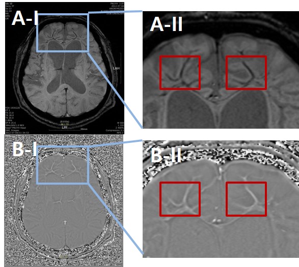
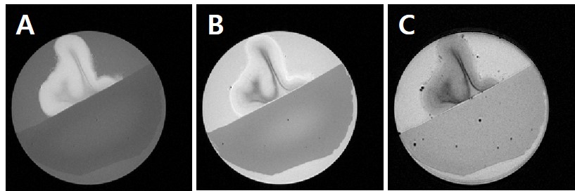
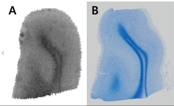
Frontal linear hypointensity and positive phase shift on in vivo SWI was accompanied by a striking frontal lobe atrophy (figure 1). Postmortem SWI revealed marked hypointensity and positive phase shift in outer WM layers beneath cortical gray matters. The LFB stained region showed the symmetrical central myelin pallor with preserved U fibers. CD68- and Ferritin- positive microglia were abundantly located in central demyelinating lesions. Ferritin immunohistochemistry also observed the white matter–cortical grey matter junction without accompanying iron content. However, the minerals, such as iron pigments and calcifications, were rarely detected. Co-registration between postmortem MRI (Figure 2) and histology demonstrated that phase contrast in subcortical WM were mainly attributed to the remnant U-fibers rather than ferritin (figure 3).Discussion
The myelin architectural changes, marked central myelin loss with preserved U-fibers beneath cortical gray matter, mainly contributed to the susceptibility contrast in frontal white matters of adult-onset leukoencephalopathy with neuroaxonal spheroids and pigmented glia (ALSP). The striking susceptibility change due to the demyelination makes the complicated susceptibility layers along the U fiber, which bring distorted main magnetic field along MRI scan and significant changes in MR signals. Because gradient echo based phase and SWI are sensitive to the susceptibility change rather than other images, they showed abnormal signal intensities at the application to ALSP cases.Acknowledgements
No acknowledgement found.References
1. Haacke, E. Mark, et al. "Characterizing iron deposition in multiple sclerosis lesions using susceptibility weighted imaging." Journal of Magnetic Resonance Imaging 29.3 (2009): 537-544.
2. Rademakers, Rosa, et al. "Mutations in the colony stimulating factor 1 receptor (CSF1R) gene cause hereditary diffuse leukoencephalopathy with spheroids." Nature genetics 44.2 (2012): 200-205.
3. Freeman, Stefanie H., et al. "Adult onset leukodystrophy with neuroaxonal spheroids: clinical, neuroimaging and neuropathologic observations." Brain pathology 19.1 (2009): 39-47.
Figures