4159
Imaging Impairment of the Glymphatic System after Diabetes1Neurology, Henry Ford Health System, Detroit, MI, United States, 2Physics, Oakland University, Rochester, MI, United States, 3Radiology, Henry Ford Health System, Detroit, MI, United States
Synopsis
The recently discovered glymphatic system has fundamentally altered the traditional model of cerebrospinal fluid (CSF) hydrodynamics and shown promising results in applications for understanding neurological diseases1, 2. However, little is known how diabetes affects the glymphatic system. The current study is the first investigation of the effect of diabetes on the glymphatic system and the relationship between glymphatic system and cognitive impairment in diabetic rats using MRI and fluorescence imaging.
Purpose
The brain lacks specialized organ-wide anatomic structure to facilitate lymphatic clearance although the brain has complex architecture and high metabolic activity. The glymphatic system has recently been developed and shown to clear brain extracellular solutes. The impairment of glymphatic system may contribute to both initiation and progression of neurological diseases1, 2. The current study is the first investigation of the effect of diabetes on the glymphatic system and the relationship between the glymphatic system and cognitive impairment in Type-2 diabetes mellitus (DM) rats using MRI and fluorescence imaging.Methods
Male Wistar rats, 13 months of age were either subjected to nicotinamide and streptozotocin induced type 2 diabetes (n=26) or without induction of diabetes (non-DM, n=18). MRI measurements were performed with a Bruker 7 T MRI. The dynamic cerebrospinal fluid (CSF) - interstitial fluid (ISF) exchange was measured continuously for 6 hours using dynamic 3D T1-weighted images with 3 baseline scans followed by intra-cisterna magna (ICM) Gd-DTPA contrast delivery via the indwelling catheter, while MRI acquisitions continued. Clearance time constant was derived using a two phase model from time evolution curve of regional tissue uptake of the paramagnetic contrast agents. To validate MRI measurements, laser scanning confocal microscopy (LSCM) images were performed 3 and 6 hours after intrathecal injection of Texas Red-conjugated dextran 3 (TR-d3). Cognitive dysfunction of animals was measured by Morris water maze and olfactory learning and memory tests.Results
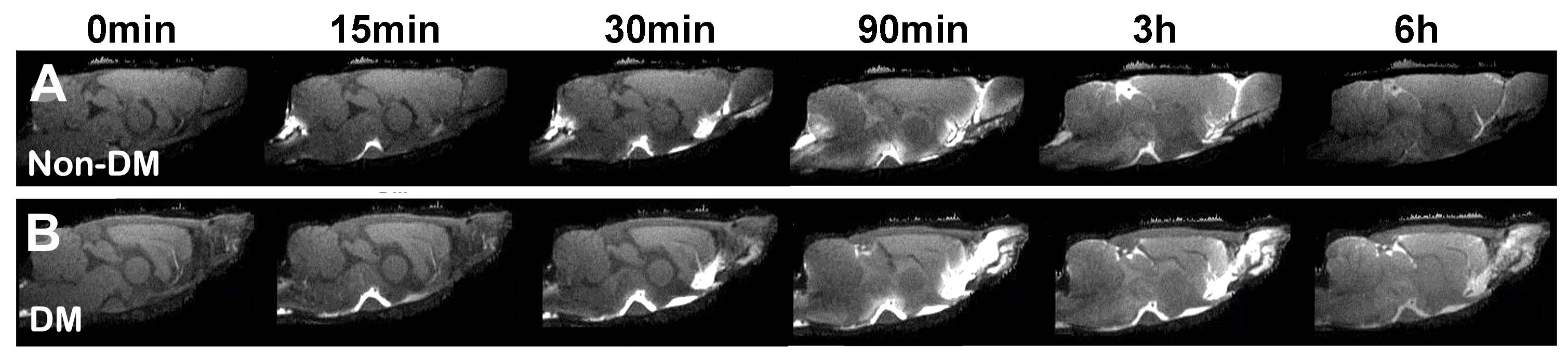
MRI CSF-ISF measurements revealed that clearance of CSF contrast agent from the interstitial space was slowed in the DM rats compared to the non-DM rats. As shown in Fig 1, compared with the non-DM rat (A), the time point matched DM rat (B) exhibits increased hyperintensity areas caused by elevated Gd-DTPA during the initial para-vascular arterial influx (15-90 min), increased the overall intensity during the middle (1.5-3 h) and end of the experiment (6 h, A vs B). Consistently, the fluorescence imaging data confirmed MRI findings of increased tracer accumulation in the areas close to para-vascular arterials during the middle of experiment (3h), and also increased tracer concentration at the end (6h) of the experiment in the DM compared with non-DM animals. The Gd-DTPA clearance rate constant was 3.4 times slower (p=0.022) in DM than in non-DM rats. Also, The DM rats exhibited significantly increased residual intensity (signal intensity at the end of experiment minus baseline intensity, p=0.005, 772% of control) in the hippocampus compared with the non-DM rat. A significant linear relationship between the accumulation of TR-d3 level and MRI residual intensity in the hippocampus 6h after ICM injection and the cognitive deficits measured by the Morris water maze (r2=0.948 in TR-d3 and r2=0.856 in MRI) and odor recognition tests (r2=0.991 in TR-d3 and r2=0. 86 in MRI) were detected.
Discussion and conclusion
Our data demonstrated for the first time, that diabetes adversely affects glymphatic system function, which is highly associated with impairment of learning and memory. DM suppresses clearance of ISF in the hippocampus, suggesting that an impairment of the glymphatic system contributes to DM-induced cognitive deficits. Whole brain MRI provides a sensitive, non-invasive tool to quantitatively evaluate CSF and ISF exchange in DM and possibly in other neurological disorders, with potential clinical application.Acknowledgements
Grant support: Supported by NIH grants R21 AG052735 and NS79612.References
1. Xie L, Kang H, Xu Q, Chen MJ, Liao Y, Thiyagarajan M, et al. Sleep drives metabolite clearance from the adult brain. Science. 2013;342:373-377.
2. Iliff JJ, Wang M, Liao Y, Plogg BA, Peng W, Gundersen GA, et al. A paravascular pathway facilitates csf flow through the brain parenchyma and the clearance of interstitial solutes, including amyloid beta. Science translational medicine. 2012;4:147ra111.
Figures