4156
Sporadic Jakob-Creutzfeld Disease quantitative diffusion profiles and resting state functional correlates1Institute of Radiology, University of Pavia, Pavia, Italy, 2Department of Neurology, University of California, San Francisco, San Francisco, CA, 3Memory and Aging Center, University of California, San Francisco, San Francisco, CA, 4Department of Radiology and Biomedical Imaging, University of California, San Francisco, San Francisco, CA, 5Department of Neuroradiology, IRCCS C. Mondino Neurological Institute, University of Pavia, Pavia, Italy, 6Department of Brain and Behavioral Sciences, University of Pavia, Pavia, Italy
Synopsis
Diffusion restriction on MRI is an important diagnostic finding in sporadic Jakob-Creutzfeld disease. We performed group-wise quantitative cortical and subcortical grey matter mean diffusivity analysis at 3.0 T and tested feasibility of single-subject diffusivity restriction maps, that, compared to group-wise analysis, can better describe variable patterns of involvement in sJCD. DMN-related regions demonstrated prominent diffusion restriction; thus DMN functional connectivity was evaluated. DMN showed a increased connectivity in sJCD, that based on cross-sectional analysis seems to decrease along with clinical worsening. Resting-state fMRI may be a promising candidate to evaluate involvement in sJCD especially during the preclinical phase.
Background and purpose
Hindered diffusion on diffusion weighted MRI is a characteristic finding in sporadic Jakob-Creutzfeld Disease (sJCD), showing high sensitivity and specificity for diagnosis. Given heterogeneous cortical and subcortical involvement across subjects, group-wise analyses may underestimate the actual brain involvement, even when quantitative assessment is performed 1–3. Despite the diverse MRI patterns of involvement, qualitative and quantitative diffusivity studies as well as our clinical experience suggest a preferential involvement of retrosplenial and parietal regions that are functionally part of the Default Mode Network. Our aims were 1) to quantify diffusion restriction in sJCD at 3.0 T and collaterally to obtain subject-specific quantitative maps of diffusivity restriction and 2) to evaluate functional correlates of hindered diffusion in sJCD by assessing functional connectivity of the default mode network at rest.Methods
37 subjects with probable or definite sJCD according to UCSF diagnostic criteria (64 ± 7.8 years, M/F=20/17) underwent a high angular resolution diffusion (HARDI) imaging acquisition at 3.0 T at University of California, San Francisco (UCSF) (2.2 mm3 isotropic voxel, b-value=2000 s/mm2, 55 diffusion gradients, 1 b0). Mean diffusivity (MD) and other DTI parameters maps were computed and mean values were extracted for 40 cortical and subcortical VOIs per side. Collaterally, voxel-wise age-corrected z-score MD maps were created for each subject, using a statistical threshold of -2 SD from average MD of controls, obtained for each lobe. A subset of 19 subjects underwent resting state fMRI acquisition protocol as well (240 volumes; field of view = 230×230mm2; matrix = 92×92; in-plane resolution = 2.5×2.5mm). In line with known involvement of parietal and retro-splenial cortices in sJCD, Default Mode Network connectivity was tested. Moreover functional data were analyzed in a pseudo-longitudinal approach using Barthel score and disease ratio (time from disease onset to MRI/total disease duration). Prion gene, PRNP, analysis, identified no mutations. Codon 129 polymorphisms were available for all subjects. Prion typing was available for the 27 subjects whom had brain autopsy. A detailed standardized neurological examination and testing including Mini-Mental State examination (MMSE), modified Barthel index, were recorded ± 3 days from MRI scan date. Thirty age- and gender-matched healthy subjects were enrolled as controls.
Results
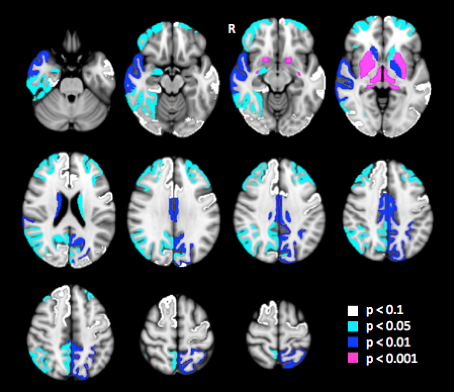
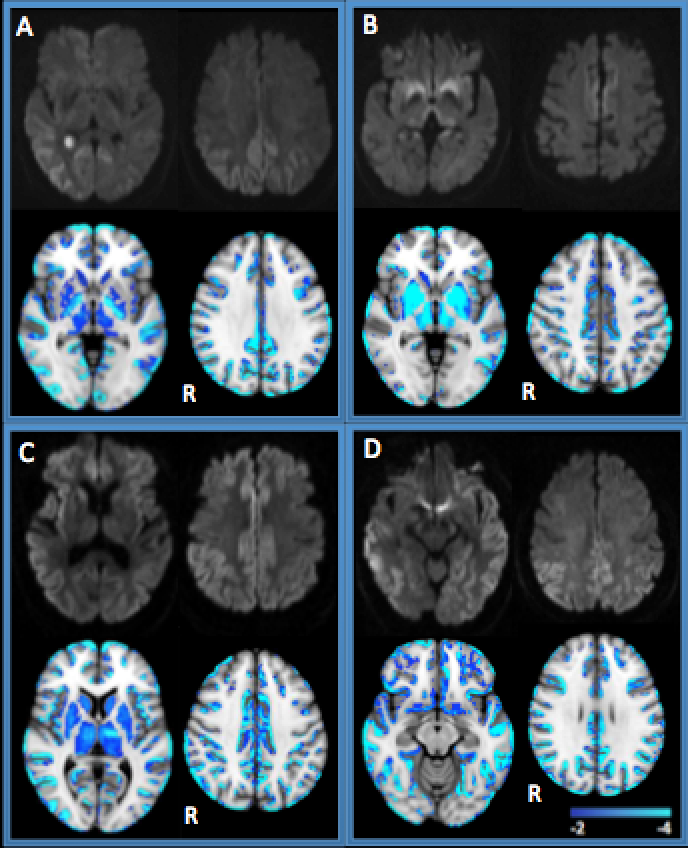
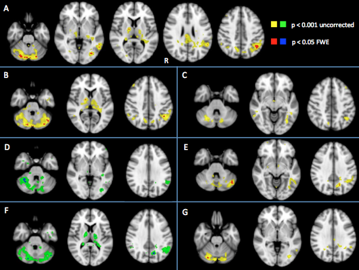
Consistent with our previous findings at 1.5 T, a significant MD reduction was shown in subcortical grey matter (p<0.0001) and in the whole cortex (p<0.05), with extended involvement of parietal, parietal-occipital and retro-splenial cortices (Figure 1). Frontal involvement was documented as well. Mean diffusivity was conversely increased in the cerebellum, coherently with previous experiences of ADC quantification 4. Single-subject voxel-wise mean diffusivity maps were created and also summed to get the involvement across the entire cohort. Although they highly correlated with visual assessment of diffusion trace images and group-wise volume-of-interest analysis results, single subjects and sum maps demonstrated more clearly areas of true diffusion restriction and ruled out areas of intrinsic signal hyperintensity on DWI (e.g. insula) (Figure 2). Abnormally increased connectivity was demonstrated for DMN both in the whole cohort and in Codon 129 polymorphism MM and MV subgroups respectively. No abnormality was evident for VV subjects, presumably for the limited number of cases. Although the analysis was cross-sectional, using Barthel score as a marker for disease severity or disease ratio, increased connectivity appeared to decrease as the disease progressed. Surprisingly functional hyperconnectivity was confirmed even in the cerebellum, where, conversely to supratentorial areas, increased mean diffusivity was demonstrated. (Figure 3)Conclusions
Mean diffusivity reduction in sJCD assessed at 3.0 T is in line with previous results obtained at 1.5 T, with prominent involvement of subcortical grey matter, retrosplenial, parietal, middle temporal and parietal-occipital cortices. Additionally current analysis show frontal involvement as well 5.
Our initial experience demonstrates feasibility of single-subject quantitative assessment of MD reduction, in a way that might overcome known visual assessment limitations and that might help in reducing underestimation of disease burden.
To our knowledge we show for the first time increased connectivity within the DMN in sJCD, confirming the hypothesis of connectivity abnormality in areas frequently involved in the disease. Functional alteration seems to be present from initial phases of the disease, suggesting a role of task-free fMRI during the prodromal state. Resting-state connectivity can be considered as a promising candidate for prion disease diagnostic assessment and follow-up. Longitudinal analysis of fMRI is currently being investigated.
Acknowledgements
No acknowledgement found.References
1. Meissner, B. et al. MRI lesion profiles in sporadic Creutzfeldt-Jakob disease. Neurology 72, 1994–2001 (2009).
2. Caverzasi, E. et al. White matter involvement in sporadic Creutzfeldt-Jakob disease. Brain 137, 3339–54 (2014).
3. Caverzasi, E. et al. Application of quantitative DTI metrics in sporadic CJD. NeuroImage Clin. 4, 426–435 (2014).
4. Cohen, O. S. et al. MRI detection of the cerebellar syndrome in Creutzfeldt-Jakob disease. Cerebellum 8, 373–381 (2009).
5. Zerr, I. et al. Updated clinical diagnostic criteria for sporadic Creutzfeldt-Jakob disease. Brain 132, 2659–2668 (2009).
Figures