4154
MR-NODDI Evaluating Amyotrophic Lateral Sclerosis: a Tract Based Spatial Statistics Analysis1Fujian Medical University Union Hospital, Fuzhou, People's Republic of China, 2Tongji Hospital, HUST, Wuhan, People's Republic of China
Synopsis
This study uses NODDI, a novel tool, to quantify changes of white matter (WM) skeleton in patients with ALS. By TBSS analysis, significant FA reductions were demonstrated within the (pre)frontal WM, partial parietal WM, corpus callosum and partial corticospinal tract. Similarly, significant Ficvf reductions were found within almost all the WM skeleton, which was more extensive than that of FA. In contrast, ODI showed no significant changes in all the WM skeleton. Therefore, NODDI is a more potential tool to demonstrating the neurite density reductions for ALS patients, which will help lead to earlier diagnosis and treatment for ALS.
Objectives
Amyotrophic lateral sclerosis (ALS) is an incredibly lethal disease and resistant to therapy. Symptoms of ALS can be similar to those of a wide variety of other, more treatable diseases or disorders. However, no test can provide a definite diagnosis of ALS at present. The aim of this study is to use neurite orientation dispersion and density imaging (NODDI), a novel tool, to quantify changes of white matter (WM) skeleton in patients with ALS compared to healthy controls.Methods
Two shells of diffusion-weighted magnetic resonance images (shell 1: b=1000s/mm2, direction=25; shell 2: b=2500s/mm2, direction=25) were acquired from 12 patients with ALS and 12 age- and sex-matched healthy controls. FMRIB's Linear Image Registration Tool was used to register all diffusion-weighted volumes to their corresponding b = 0 s/mm2 volume, and to correct for motion and eddy currents. Subsequently, the Brain Extraction Tool was used to remove non-brain tissue, FSL's DTIFIT was used to calculate maps of FA, and NODDI toolbox was used to calculate the intra-neurite volume fraction (Ficvf) indicating the neurite density and orientation dispersion index (ODI). FSL's Tract-Based Spatial Statistics (TBSS) tool was used to align individual FA maps to FSL’s standard adult FA template. Following registration, the FA maps of all subjects were thinned to create white matter skeletons. Then, Ficvf and ODI maps were created and registered using the TBSS registrations of FA to the adult FA template, and the skeleton mask was applied to the registered images. The white matter skeletons differences in FA, Ficvf and ODI were compared between ALS patients and healthy controls.Results
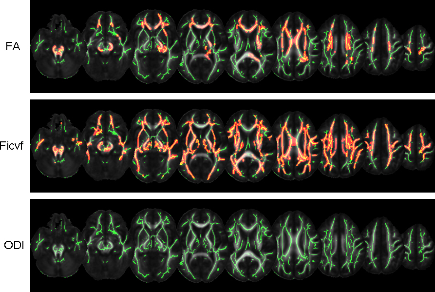
In the TBSS analysis, significant FA reductions were demonstrated within the (pre)frontal WM, partial parietal WM, corpus callosum and partial corticospinal tract (with TFCE-correction). Similarly, significant Ficvf reductions were found within almost all the WM skeleton (with TFCE-correction), which was more extensive than that of FA. In contrast, ODI showed no significant changes in all the WM skeleton.Discussion
ALS commonly affects the upper motor neurons in the brain or part of the brain stem first, and in later phase, the disease will spread and affect other regions of the brain. Therefore, significant Ficvf reductions can be observed within almost all the WM skeleton, which indicates the reductions of the neurite density in the corresponding WM. Ficvf found more extensive changes than FA, indicating Ficvf is a more sensitive parameter. Although neurite density is different between ALS patients and healthy controls, the neurite orientation is almost the same in the WM area, indicating low neurite orientation dispersion in both groups. Therefore, ODI showed no significant changes in the WM skeleton.Conclusion
NODDI is a more potential tool to demonstrating the neurite density reductions for ALS patients, which will help lead to earlier diagnosis and treatment for ALS.Keywords
NODDI, ALS, diagnosis, TBSS.Acknowledgements
The authors thank all the patients joining in our study and thank Chanchan Liu for his help with the ALSFRS-R evaluation.References
1. Zhang, H., et al., 2012. NODDI: practical in vivo neurite orientation dispersion and densityimaging of the human brain. NeuroImage 61 (4), 1000–1016.
2. Tariq M, Schneider T, Alexander D, Wheeler-Kingshot CAM, Zhang H. Assessing scan-rescan reproducibility of the parameter estimates from NODDI. Proceedings of the International Society for MagneticResonance in Medicine, Salt Lake City, UT; 2013.
Figures