4152
Altered White Matter Microstructure in Middle-Aged Type 2 Diabetic Patients: A Diffusional Kurtosis Imaging Study Based on Two-compartment White Matter Model1Shaanxi Provincial People's Hospital, Xi'an, People's Republic of China, 2Clinical science, Philips Healthcare China
Synopsis
This study aims to use a two-compartment diffusion model of white matter based on diffusional kurtosis imaging (DKI) to explore early white matter alternations in middle-aged type 2 diabetes mellitus (T2DM) . 33 T2DM patients and 13 healthy control were enrolled. All diffusion parameters (FA=fractional anisotropy, MD=mean diffusivity, AD=axial diffusivity, RD=radial diffusivity, MK=mean kurtosis, AK=axial kurtosis, RK=radial kurtosis, Da=intra-axonal diffusivity, De∥=axial extra-axonal space diffusivity, De⊥= radial extra-axonal space diffusivity) were compared. De⊥ was demonstrated to be the most sensitive in detecting the diffusion changes. These increased De⊥ (extra-axonal diffusivity) and unchanged Da (intra-axonal diffusivity) reflected the increased water and/or demyelination.
Introduction
Type 2 diabetes mellitus (T2DM) has emerged as an important risk factor for cognitive impairment and dementia1-2. Early detection of brain abnormalities at the preclinical stage can be useful for developing preventive interventions to abate cognitive decline. Previous diffusion tensor imaging (DTI) studies have revealed widespread white matter (WM) alternations in elderly patients. Diffusional kurtosis imaging (DKI) is a clinically feasible extension of DTI that examines the additional contribution of non-Gaussian diffusion effects as a result of brain microstructural complexity. Jensen et.al proposed a two-compartment non-exchange diffusion model of WM based on DKI analysis and provides analytical expressions for the intra- and extra-axonal diffusion tensors3. So this study aims to further explore early white matter changes in middle-aged T2DM.Methods
This study was approved by the local institutional review board. Subjects: 33 T2DM patients (DM group; based on diagnostic criteria of American Diabetes Association; 56.21±6.28 years old, 10 females) and 13 healthy control (HC group, 54.74±5.74 years old, 4 females) who underwent MRI were enrolled. A battery of neuropsychological tests including Montreal Cognitive Assessment and Mini-Mental State Examination were performed at first. MRI acquisition: Conventional MRI and DKI were performed on a 3.0T scanner (ingenia, Philips Medical Systems, The Netherlands). DKI protocols were: 32 directions, b value=0, 1000, 2000 s/mm2, TR/TE=6000/150ms, slice thickness= 6 mm, field of view = 224mm×224mm, matrix = 112×112, NEX = 1. Image analysis: All DKI data were processed using a custom-written program in MATLAB, and all parameters (FA=fractional anisotropy, MD=mean diffusivity, AD=axial diffusivity, RD=radial diffusivity, MK=mean kurtosis, AK=axial kurtosis, RK=radial kurtosis, Da=intra-axonal diffusivity, De∥=axial extra-axonal space diffusivity, De⊥= radial extra-axonal space diffusivity) were generated. FMRIB’s Software Library (FSL) with tract-based spatial statistics (TBSS, part of FSL)4 was used to analyze all above diffusional metrics and compare group difference with age, gender as covariates. The difference of neuropsychological scores between groups were also analyzed. All tests were taken to be significant at P<0.05.Results
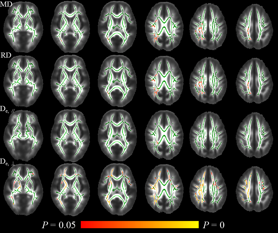
The neuropsychological scores between DM and HC group showed no significant difference. In TBSS analysis, DM group exhibited significant increase in multiple WM regions on MD, RD, De∥ and De⊥ maps (as shown in Fig.1), which involved 5.26%, 2.82%, 0.46% and 20.34% of mean WM skeleton respectively. De⊥ detected most WM changes, which mainly located in right internal capsule, external capsule, corona radiata, superior longitudinal fasciculus and bilateral frontal WM. Whereas, FA, AD, MK, AK, RK and Da showed no significant difference between two groups.Discussion
In the present study, multiple diffusional metrics were employed to detected WM microstructural changes in middle-aged T2DM patients. According to our results, De∥ seemed to be the most sensitive metric in revealing WM disruptions, which potentially provided valuable information to study diabetic encephalopathy and predict cognitive impairment. Furthermore, although many DTI studies has studied T2DM related WM alterations for a few years, there is still little study focused on middle-aged T2DM patients. Compared with previous DTI studies in elderly T2DM patients, this study demonstrated early WM impairment in right internal capsule, external capsule, corona radiata, superior longitudinal fasciculus and bilateral anterior thalamic radiations, which might reflected more vulnerable regions playing particularly important roles in T2DM-induced cognitive dysfunction4. The increased De∥, De⊥ and no significantly changed Da indicated an increased diffusivity mainly from extra-axonal space, which might be due to the increased water and/or demyelination, rather than injuries to axons.Conclusion
By using the two-compartment diffusion model based on DKI, this study demonstrated that De⊥ is more sensitive than DTI metrics in detecting the diffusion changes in middle-aged T2DM patients. These increased extra-axonal diffusivity and unchanged intra-axonal diffusivity reflected the increased water in extra-axonal space and/or demyelination, rather than injuries to axons, which provided novel insights into the possible pathological changes underlying white matter degeneration in T2DM.Acknowledgements
This work was supported by National Natural Science Foundation of China (No. 81270416). We are grateful to Dr Qin Zhang in the Endocrinology Department for the patient recruitment, and would like to thank Philips Applied Science Lab for their technical assistance. Finally, we thank all participants and their parents for their loyalty and cooperation.References
1. Crane PK, Walker R, Larson EB. Glucose levels and risk of dementia. The New England journal of medicine. 2013;369(19):1863-1864.
2. Zhang J, Wang Y, Wang J, et al. White matter integrity disruptions associated with cognitive impairments in type 2 diabetic patients. Diabetes. 2014;63(11):3596-3605.
3. Fieremans E, Jensen JH, Helpern JA. White matter characterization with diffusional kurtosis imaging. Neuroimage. 2011;58(1):177-188.
4. Smith SM, Jenkinson M, Johansen-Berg H, et al. Tract-based spatial statistics: voxelwise analysis of multi-subject diffusion data. Neuroimage. 2006;31(4):1487-1505.
Figures