4151
Brain gray matter changes in type 2 diabetes mellitus: A meta-analysis of whole-brain voxel-based morphometry studylin lin1 and Guangyao wu1
1radiology, Affiliated Zhongshan Hospital of Dalian University, Dalian, People's Republic of China
Synopsis
To our knowledge, this is the first whole-brain VBM meta-analysis showing a reduced volume of whole and regional gray matter (GM) in the brains of type 2 diabetes mellitus (T2DM) patients. we used a new meta-analytic tool, signed differential mapping to identify consistent results about global and regional abnormalities in T2DM, and explore the relationship between cognitive and GM alternations. We showed educed volume of whole and regional GM in T2DM patients, particularly in the temporal lobe, the GM volumes of the right insula were positively correlated with MMSE scores, and those changes may indicate a risk of dementia.
Purpose
Increasing neuroimaging studies of voxel-based morphometry (VBM) have revealed gray matter (GM) anomalies in patients with type 2 diabetes mellitus (T2DM)1. However, not all the studies reported entirely consistent findings, which may be due to small and heterogeneous samples of participants2-4, there has been increasing interest in using the meta-analysis approach to identify consistent results. So the aim of our study was to identify the alternations of global gray matter volumes (GMV) and consistent regional abnormalities in T2DM patients.Method
A systematic search of relevant studies published in PubMed and Embase databases from January 2000 to March 2016 was conducted using a combination of keywords: (“voxel-based” OR “voxel-wise” OR “volumetric” OR “voxel based morphometry” OR “VBM” OR “morphometry” OR “gray matter”) AND (“diabetes” OR “diabetic” OR “diabetes mellitus” OR “type 2 diabetes mellitus” OR “T2DM”). Neuroimaging studies of both volumetric data and whole-brain VBM data were included. A quantitative meta-analysis of volumetric and whole-brain VBM studies in patients with T2DM compared with healthy controls (HCs) were performed by means of STATA package v.12.0 and anisotropic effect size version of signed differential mapping (AES-SDM) software package respectively.Result
A total of 15 volumetric studies(1561 T2DM patients and 869 HCs) and 5 VBM studies (110 T2DM patients and 103 HCs) on GM investigations in T2DM patients vs. HCs were identified. The volumetric meta-analysis showed that there were significant differences (SMD=−0.56,95%CI=−0.81 to −0.31, P<0.01) of GMV between the two groups, suggesting that GMV of patients with T2DM was lower than HCs. The whole-brain VBM meta-analysis revealed that gray matter reductions in the left superior temporal gyrus, the right middle temporal gyrus, the right rolandic operculum, and the left fusiform gyrus in the T2DM patients compared with the HCs. No GMV increases were found. The results remained largely unchanged in the following jackknife sensitivity analyses. Meta-regression analysis showed that Mini-Mental State Examination (MMSE) scores have a positive relationship with the GMV in the right insula.Discussion
We have revealed widespread GM atrophy, as well as regional atrophy, particularly in the right MTG. The MTG is implicated in neurodegeneration in T2DM such as regional neuronal loss, vascular abnormalities, micro-infarcts, hypometabolism, lower cerebral blood flow, and the like1,4. The MTG is part of the default mode network (DMN), which has been closely linked to cognition functions5. Based on these findings, we suggest that MTG atrophy may relate to cognitive dysfunction, and accelerate the transition from normal cognition to MCI in T2DM patients. The fusiform gyri are frequently identified a "visual-form area"6, our results show that The fusiform gyri is atropht inT2DM patients, which suggests that T2DM may have attention, memory and visual dysfunction. Another finding in our meta-analysis was that MMSE scores had a positive relationship with GMV in the right insula. The insula plays important integrative roles, it is also a core node of the salience network which is damaged in AD and MCI7-8Conclusion
To our knowledge, this is the first whole-brain VBM meta-analysis showing the reduced volume of whole and regional gray matter in brain of T2DM patients, especially in the temporal lobe; the GM volumes of right insula had a positive relationship with MMSE scores, and those changes may be risk for the dementia. Further longitudinal search is need to confirm the gray matters changes and cognition dysfunction and their relationship to understand the pathophysiological mechanism underlying T2DM-induced cognitive impairment.Acknowledgements
No acknowledgement found.References
1. Chen Z, Li L, Sun J, Ma L, et al. Mapping the brain in type II diabetes: Voxel-based morphometry using DARTEL. European Journal of Radiology.2012;81:1870–1876. 2. Yau PL, Javier DC, Ryan CM, Tsui WH, Ardekani BA, Ten S, Convit A,et al. Preliminary evidence for brain complications in obese adolescents with type 2 diabetes mellitus.Diabetologia.2010;53:2298–2306. 3. Zhang Y, Zhang X, Zhang J, Liu C, Yuan Q, Yin X, Wei L, Cui J, Tao R, Wei P, Wang J, et al. Gray matter volume abnormalities in type 2 diabetes mellitus with and without mild cognitive impairment. Neuroscience Letters.2014;562:1–6. 4. Natalia Garc´ia-Casares, Marcelo L. Berthier, et al. Structural and Functional Brain Changes in Middle-Aged Type 2 Diabetic Patients: A Cross-Sectional Study. Journal of Alzheimer's Disease.2014;40:375–386. 5. Fox MD, Corbetta M, Snyder AZ, Vincent JL, Raichle ME, et al.Spontaneous neuronal activity distinguishes human dorsal and ventral attention systems. Proc.Natl. Acad. Sci. U. S. A.2006;103: 10046–10051. 6. Dehaene, S., Cohen, L, et al. The unique role of the visual word form area in reading. Trends CognSci.2011;15:254–262. 7. Kurth Florian, Zilles Karl, Peter T. Fox, Angela R. Laird, Simon B. Eickhoff. A link between the systems: functional differentiation and integration within the human insula revealed by meta-analysis. Brain StructFunct .2010;214:519–534. 8. Menon V,Uddin LQ. Saliency, switching, attention and control: a network model of insula function. Brain StructFunct. 2010;214: 655-667.Figures
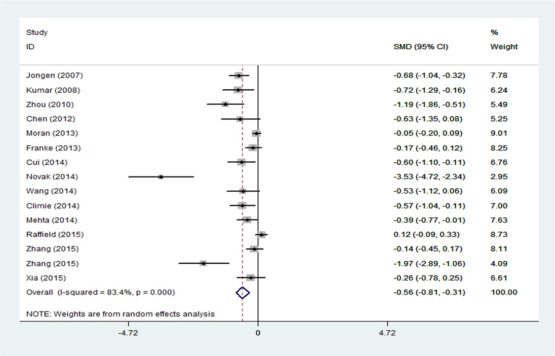
Forest plot of global
GMV decreases in patients with T2DM compared with HCs.
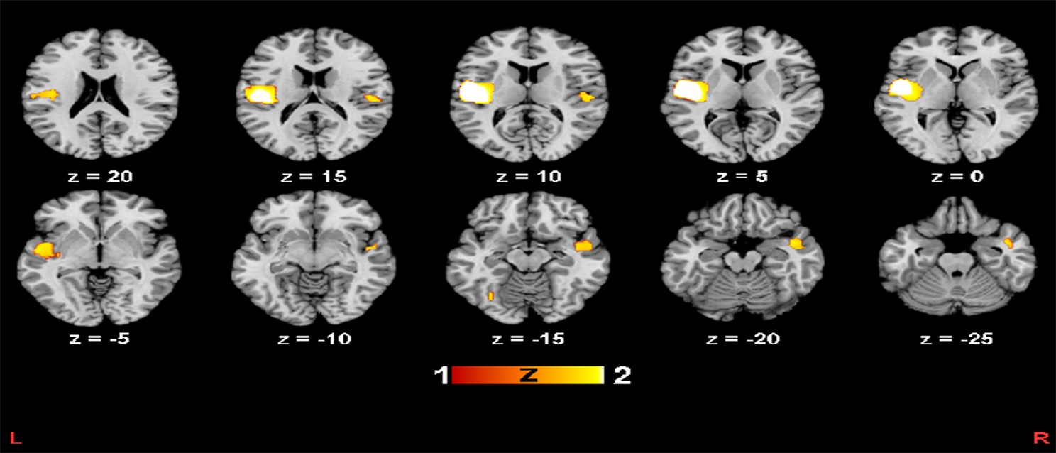
GMV decreases in
patients with T2DM compared with HCs.
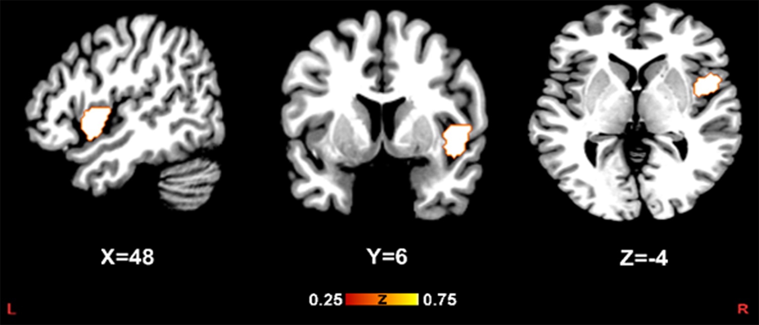
Results
of the meta-regression analysis showing a significant association between MMSE
scores and GMV in the right insula.