4147
Altered Topological Properties of Structural Connectome in Early-Stage, Drug-naive PD Revealed by Graph Theoretical Analysis1Radiology, West china hospital, Chengdu, People's Republic of China, 2Psychosis Studies, Institute of Psychiatry, Psychology & Neuroscience, King’s College London, United Kingdom, 3Neurology, West china hospital, People's Republic of China, 4Medical Information Engineering, School of Electrical Engineering and Information, Sichuan University, People's Republic of China, 5Radiology, Henan Provincial People's Hospital & the People's Hospital of Zhengzhou University, People's Republic of China
Synopsis
To use diffusion tensor imaging (DTI) and graph theory approaches to explore the brain structural connectome in patients with early-stage drug-naive Parkinson's disease (PD). The structural connectome was constructed by using deterministic tractography and thresholding the mean fractional anisotropy of 90 brain regions to yield 90*90 partial correlation matrixes. The decreased characteristic path length Lp and increased global efficiency Eglob in the PD patients relative to healthy controls indicated that structural networks are closer to randomization, providing a structural basis for functional alterations of PD.
Purpose
Parkinson's disease (PD) is a common degenerative disorder of the central nervous system. The underlying pathological process is not confined to the nigrostriatal pathway, also involving widespread cerebral cortical areas1. Previous neuroimaging studies have investigated the brain functional connectome of PD, but it is unclear to what extent structural abnormalities underlie functional alterations. This study aims to explore the topological organization of whole-brain structral network in patients with early-stage drug-naive PD.
Methods
Magnetic resonance (MR) scanning was carried out in Trio Tim (3T) MR imaging system (Siemens; Erlangen). Diffusion tensor imaging (DTI) and high resolution T1-weighted images were obtained from 41 early-stage drug naïve PD patients and 35 age- and sex-matched healthy controls (HC). Their brain images were segmented into 90 regions using automated anatomic labeling (AAL)2 atlas. Structural connectivity between these regions was established using partial correlation coefficient. Whole-brain structural connectome was constructed by thresholding the resultant partial correlation matrix (90*90). Graph theory analysis3 was then employed to examine group specific topological characteristics. Nonparametric permutation test was used for multiple comparison correction. In addition, between-group differences and relationships with Unified PD Rating Scale part III (UPDRS-3) which was used to assess the motor disability were investigated.Results
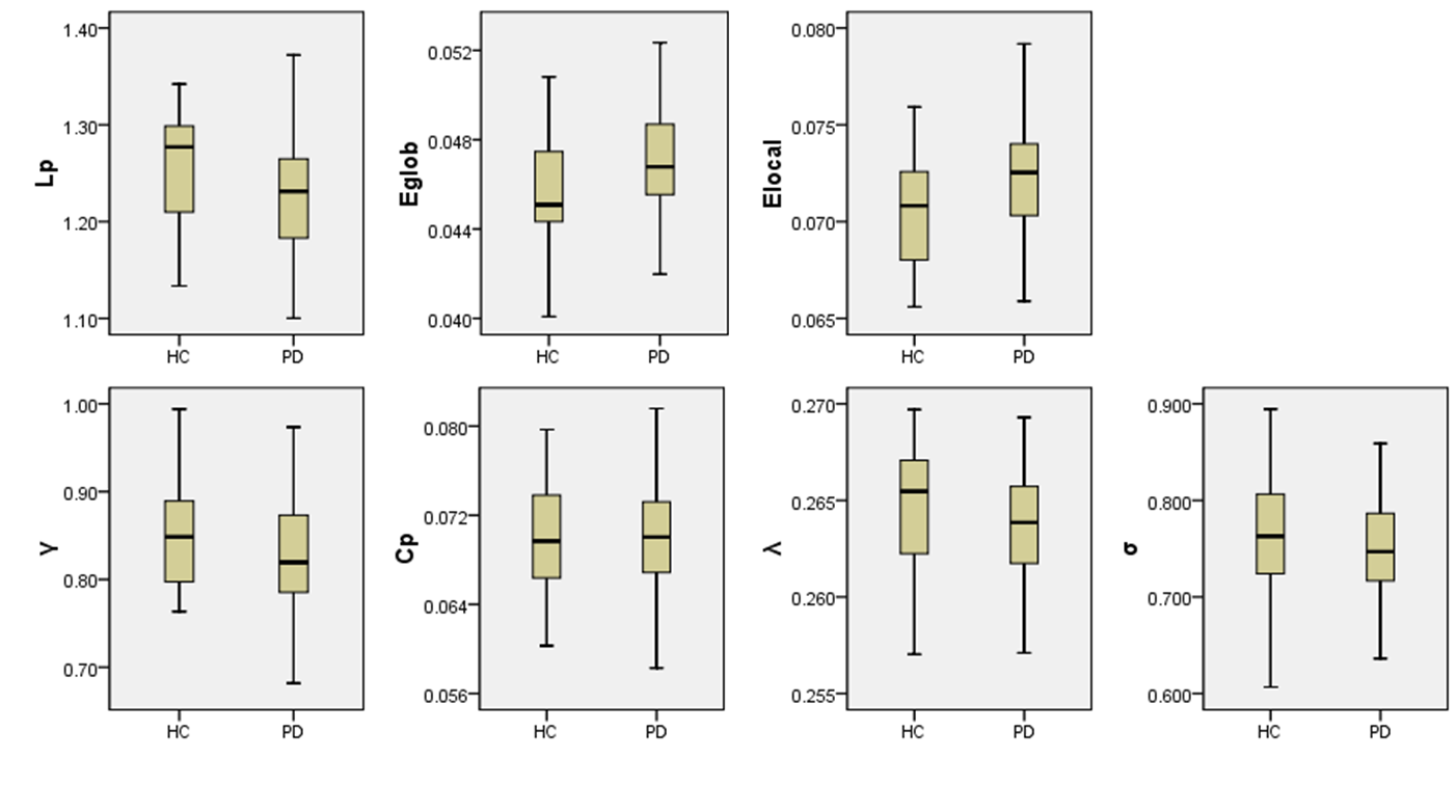
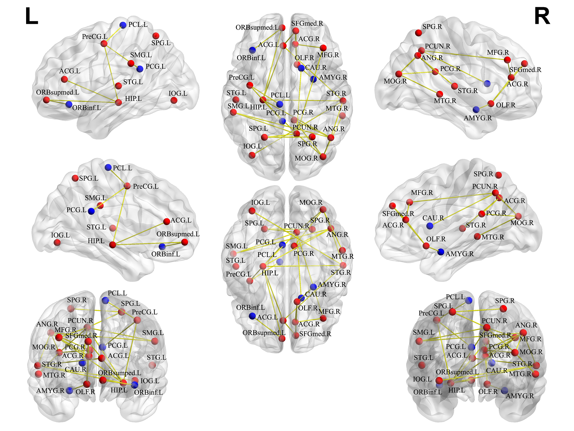
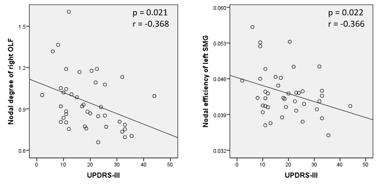
Both the PD and HC groups showed small-world architecture in brain structral networks. However, the PD patients had significantly decreased characteristic path length Lp (P = 0.0114), and increased global efficiency Eglob (P = 0.0076) and local efficiency Elocal (P = 0.0118), with no significant differences in clustering coefficient Cp (P = 0.9156), normalized clustering coefficient γ (P = 0.2605), normalized characteristic path length λ (P = 0.5457) or small-worldness σ (P = 0.2478) (Figure 1). At nodal level, decreased nodal centralities were found in striatum-limbic-frontal regions, and increased mainly in default mode network, frontal-parietal network and visual cortex of PD patients (Figure 2). The nodal degree of right olfactory cortex and nodal efficiency of left supramarginal gyrus was negatively correlated with Unified PD Rating Scale (UPDRS) part III score (Figure 3).
Discussion
This study applied graph analysis combined with DTI to assess large-scale structural networks in early-stage drug-naive PD patients. Compared with HC, the decreased Lp and increased Eglob in the PD patients indicated that structural networks are closer to randomization which was in line with the previous functional connectome study in PD4. Furthermore, PD patients showed decreased nodal centralities in the striatum-limbic-motor regions, which was in accordance with the classical emotion and motor impairment. Increased nodal characteristics in default mode network, frontal-parietal network and visual cortex might reflect compensatory changes in response to motor and visual impairment. Specifically, asymmetric alterations in the posterior cingulate cortex (PCC) appears in PD versus HC as decreased nodal centrality in left PCC and increased nodal centrality in right PCC. This effect represents abnormally low inter-hemispheric PCC correlations. The nodal degree of right olfactory cortex and nodal efficiency of left supramarginal gyrus was negatively correlated with UPDRS-III score, which were associated with olfactory dysfunction and motor impairment in PD.Conclusion
The structural connectome in early-stage drug naïve PD patients showed a shift toward ‘randomization’, providing a structural basis for functional alterations of PD. The abnormalities in widespread brain regions were compatible with the current notion that PD can be understood by investigating the dysfunction of large-scale, spatially distributed neural networks. This study demonstrates a disruption of whole-brain topological organization of the brain structural connectome in early-stage drug-naive PD patients.Acknowledgements
This study was supported by the National Natural Science Foundation (Grant Nos. 81621003, 81220108013, 81171488, 81227002, 81030027, 81171286, 81501452 and 30830046), the Program for Changjiang Scholars and Innovative Research Team in University (PCSIRT, grant IRT1272) of China, the Changjiang Scholar Professorship Award (Award No. T2014190) of China, and the CMB Distinguished a Professorship Award (Award No. F510000/ G16916411) administered by the Institute of International Education.References
1. Zhang J, Bi W, Zhang Y, et al. Abnormal functional connectivity density in Parkinson's disease. Behav Brain Res.2015; 280:113-118
2.Tzourio-Mazoyer N, Landeau B, Papathanassiou D, et al. Automated anatomical labeling of activations in SPM using a macroscopic anatomical parcellation of the MNI MRI single-subject brain. NeuroImage. 2002; 15:273–289.
3.Bullmore E, Sporns O. Complex brain networks: Graph theoretical analysis of structural and functional systems. Nat Rev Neurosci. 2009; 10:186–198.
4. Luo CY, Guo XY, Song W, et al. Functional connectome assessed using graph theory in drug-naive Parkinson's disease. J Neurol. 2015;262(6):1557-67.
Figures