4128
The disturbed subcortical local intrinsic activity synchronism in mild cognitive impairment and its association with spatial navigation ability: a resting-state fMRI study1Department of Radiology, Affiliated Drum Tower Hospital of Nanjing University Medical School, Nanjing, China, NanJing, People's Republic of China, 2Department of Neurology, Affiliated Drum Tower Hospital of Nanjing University Medical School, Nanjing, China, NanJing, People's Republic of China, 3Memory Disorders Clinic, Department of Neurology, Charles University in Prague, 2nd Faculty of Medicine and Motol University Hospital, Prague, Czech Republic, Czech Republic, 4International Clinical Research Center, St.Anne’s University Hospital Brno, Brno, Czech Republic, Czech Republic, 5Philips Healthcare, Shanghai, People's Republic of China, People's Republic of China, 6Philips Healthcare, HongKong, People's Republic of China, People's Republic of China
Synopsis
Loss of spatial navigation skills is a typical feature in mild cognitive impairment. Here we investigate intrinsic activity using resting-stat functional magnetic resonance imaging within the subcortical regions, which is previously reported to be important in spatial navigation. Right hippocampus, pallidum and thalamus showed significant decreased regional homogeneity of local intrinsic activity in the patients, and the correlational trend between regional homogeneity and allocentric navigation performance showed significantly difference between patients and normal aging controls. These results showed evidences for the intrinsic subcortical activity damage and a possible compensatory mechanism of spatial navigation in the early mild cognitive impairment.
Purpose
Loss of spatial navigation skills is a typical feature in the early stage Alzheimer disease (AD) and mild cognitive impairment (MCI), and seriously affects the patients’ life quality. Therefore, exploration of neural mechanism of such navigation impairment is a particularly important issue in both medicine and neuroscience. Specifically, some subcortical regions (e.g. hippocampus, entorhinal cortex) have been reported to play a critical role in the spatial navigation1,2. Putatively, the functional disorder of these regions in AD/MCI patients may be responsible to their impairment of navigation. Particularly, the intrinsic activity has been supposed as an important model of human brain activity, and resting-state fMRI has been widely and successfully used to detect the disorder of intrinsic activity, such like in AD/MCI patients3. Therefore, it is intriguingly to investigate that if and how the spatial navigation impairment is associated with an abnormal intrinsic activity within those subcortical regions.Methods
30 MCI
patients and 30 sex, education, age-matched participants were recruited from
the neurology department of Nanjing Drum Tower Hospital. The NINCDS-ADRDA was
used as diagnosis criteria. Written consent was obtained from all subjects and
there is no history of major neurological or psychiatric disorder in normal
control elderly.
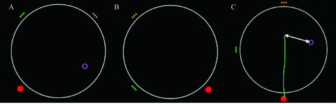
A spatial
navigation task was used to evaluate the navigation performance for all the
participants, which is a computer simulation task, following a previous study4. Specifically, there were three parts: 1) allocentric-egocentric
navigation, 2) egocentric navigation and 3) allocentric navigation, each of
which was repeated for eight times. The total error of the eight repeat was
recorded as an evaluation of the over-all, egocentric and allocentric
navigation ability (Figure 1).
Both T1
weighted and BOLD functional magnetic resonance image (MRI) data was acquired in
the same hospital at two Philips 3T MR Scanners (Achieva TX and Ingenia, Best,
the Netherlands). The T1 data was first used to identify 14 subcortical regions
(bilateral pallidum, putamen, thalamus, hippocampus, amygdala, accumbens,
caudate) using the FIRST program in FSL. Then the T1 image was coregistered
with the functional MRI image. Three typical local intrinsic activity measures,
namely, amplitude of low frequency fluctuation (ALFF)5, fractional ALFF (fALFF)6 and
regional homogeneity (ReHo)7, was then calculated to estimate the local intrinsic activity level
within the 14 subcortical regions.
A two sample t-test
was first applied on ALFF, fALFF and ReHo value in all the subcortical regions between
MCI and NC groups. For those measures and regions showing significant group
difference, a correlational analysis was then performed between the spatial
navigation performances and fMRI measures.Results
Intriguingly,
only egocentric spatial navigation performances showed significant difference between
MCI and NC groups (Allo-Ego, T=1.35, p=0.19; Allo, T=0.47, p-0.64; Ego, T=2.12,
p=0.04).
After controlled
age, gender, education history, and scanner type as covariates, ALFF and fALFF
showed no significant group difference within any of the subcortical regions.
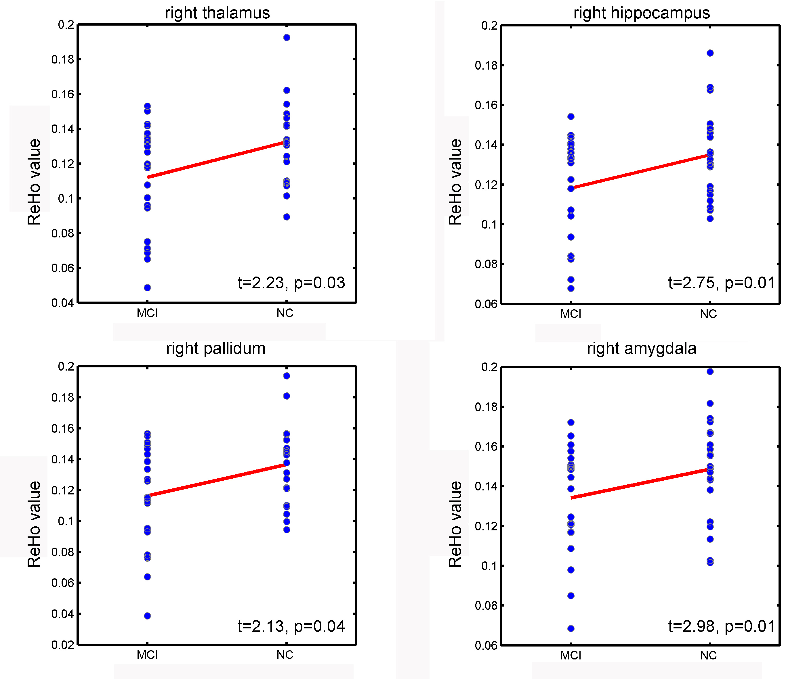
However, in right thalamus, hippocampus, pallidum and amygdala, the ReHo value
were significantly decreased in MCI group (Thalamus, T=2.24, p=0.03; Pallidum,
T=2.13, p=0.04; hippocampus, T=2.75, p=0.01; amygdala, T=2.98, p=0.01; Figure 2).
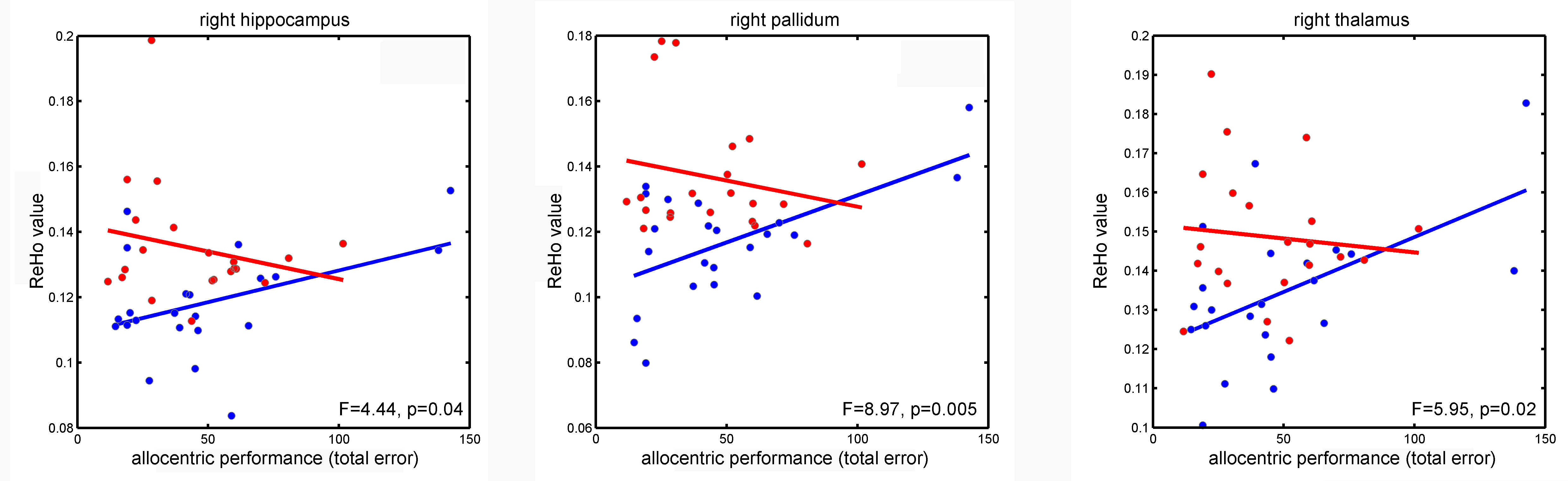
The
correlational analysis between ReHo and spatial navigation performance within
the four regions mentioned above showed very intriguing results. ReHo value and
navigation performances showed no significant correlation within each of the groups.
However, the correlational trend between ReHo and allocentirc spatial
navigation showed a significant difference between MCI and NC groups in right hippocampus
(F=4.44, p=0.04), thalamus (F=5.95, p=0.02) and pallidum (F=4.28 p=0.05), which
was positive in NC group and negative in MCI group (Figure 3)Discussion
In the current study, we found ReHo value is significant decreased within hippocampus, palladium, amygdala and thalamus regions in the MCI group. This local signal de-synchronism in the MCI patients indicated that local neural functional disorder within such regions. These regions like hippocampus is previously reported to played critical role in allocentric navigation1,2. Here, the allocentric navigation performance tend to positively correlated with ReHo value within hippocampus, pallidum and thalamus in NC group, but negatively correlated with ReHo in MCI group. Therefore, given that the allocentric performance is not significantly decreased, it is possible that these ReHo reduction and interaction effects indicated a compensatory mechanism in the early stage of dementia in human brains. Moreover, the egocentric navigation loss was found in our data, but no brain-behavior association was detected. It may related to other brain regions and need further investigation. In summary, our study found evidences of altered subcortical intrinsic activity in MCI and its association with navigation performance, which may provide a possible model of the mechanism underlying the navigation impairment in MCI.Acknowledgements
No acknowledgement found.References
1. Moffat, S.D., W. Elkins, and S.M. Resnick, Age differences in the neural systems supporting human allocentric spatial navigation. Neurobiol Aging, 2006. 27(7): p. 965-72.
2. Nedelska, Z., et al., Spatial navigation impairment is proportional to right hippocampal volume. Proc Natl Acad Sci U S A, 2012. 109(7): p. 2590-4.
3. Zhang, D. and M.E. Raichle, Disease and the brain's dark energy. Nat Rev Neurol, 2010. 6(1): p. 15-28.
4. Hort, J., et al., Spatial navigation deficit in amnestic mild cognitive impairment. Proc Natl Acad Sci U S A, 2007. 104(10): p. 4042-7.
5. Zang, Y.F., et al., Altered baseline brain activity in children with ADHD revealed by resting-state functional MRI. Brain Dev, 2007. 29(2): p. 83-91.
6. Zou, Q.H., et al., An improved approach to detection of amplitude of low-frequency fluctuation (ALFF) for resting-state fMRI: fractional ALFF. J Neurosci Methods, 2008. 172(1): p. 137-41.
7. Zang, Y., et al., Regional homogeneity approach to fMRI data analysis. Neuroimage, 2004. 22(1): p. 394-400.
Figures