4126
The effects of a Western-type diet on the cerebrovascular response to hypercapnia in a double transgenic mouse model for Alzheimer's DiseaseKristof Govaerts1, Jessica Sternisa1, Tom Dresselaers2, Fred Van Leuven3, and Uwe Himmelreich1
1Imaging & Pathology, KU Leuven, Leuven, Belgium, 2Radiology, UZ Leuven, Leuven, Belgium, 3Human Genetics, KU Leuven, Leuven, Belgium
Synopsis
Alzheimer's Disease (AD) is the most common neurodegenerative disease, and is influenced by various environmental factors. In this study, we evaluate the effect of a high-fat, high-sugar ‘Western-type’ diet on the on the vascular response capacity to hypercapnia in a double transgenic (APP/PS1) mouse model of AD. We make use of Arterial Spin Labelling to investigate basal perfusion and cerebrovascular response to hypercapnia (CVR) in the hippocampus, cortex and thalamus.
Target audience
This abstract is relevant to researchers interested in the effect of diet on the development and progression of Alzheimer's Disease, as well as those interested in arterial spin labelling, vascular response and transgenic mouse models.Purpose
Alzheimer's Disease (AD) is the most common neurodegenerative disease. During recent years, the crucial role of vascular risk factors has been brought to the foreground of AD research1, indicating that lifestyle plays an important role in the development of AD. Epidemiological data suggests that diets high in saturated fats, which are common in the Western world, can reduce cardiovascular health and may therefore influence the onset of AD2. We hypothesized that a ‘Western-type diet’, high in saturated fats, sugar and cholesterol, could influence pathology in a transgenic mouse model for AD. The readout we examined was the cerebrovascular response to hypercapnia (CVR), as we have previously described this to be a sensitive early marker in a different AD model3.Methods
We analyzed male double transgenic APP.V717IxPS1.A246E mice4, denoted APP/PS1 (n=7) and their age-matched nontransgenic controls (n=7). After the initial scanning session at 2mo, these groups were subdivided into Western-type diet (TD88137, ssniff Spezialdiäten, ‘WD’, n=4/4) and control diet (CD88137, ssniff Spezialdiäten, ‘CD’, n=3/3) cohorts. Animals were anesthetized using a mixture of 150 mg/kg ketamine (Anesketin, Eurovet, Bladel, NL), 3.8 mg/kg midazolam (Dormicum, Roche, Brussels, BE) and 0.5 mg/kg atropine (Atropine sulfate, Sterop, Brussels, BE) intraperitoneally (i.p.), divided over two to three injections over 20 minutes. Animals were subsequently intubated and mechanically ventilated with pure oxygen as described previously5, followed by an i.p. injection of 8 mg/kg rocuronium bromide (Esmeron, Organon, Oss, NL) for respiratory muscle paralysis. Expired CO2 values were continuously monitored (Vaisala Carbocap Carbon dioxide transmitters series, Bonn, GE), as well as body temperature and respiratory and heart rates (SAII, Stony Brook, NY, USA). The hypoventilation challenge was achieved by reducing the respiratory rate and tidal volume by 25% and 20% respectively. MR measurements were performed on a 9.4T Biospin small animal MR system (20cm horizontal bore, Bruker Biospin, Ettlingen, GE), using a 7 cm linearly polarized resonator for transmission and an actively-decoupled mouse brain surface coil for receiving (Rapid Biomedical, Rimpar, GE). ASL data were acquired using a FAIR approach6, and a RARE readout with the following specific parameters: TR 18s, TE 5.2 ms, rare factor 72, FOV 2.5x2.5 cm, matrix 128x128 with partial FT acceleration to 128x72, fourteen inversion times from 300-4000ms, using an inversion hyperbolic secant of 14ms, (Paravison 5.1, Bruker). CBF values were calculated using the T1 difference method and assuming an arterial T1 of 2400ms. CVR values were calculated by taking the ratio of the relative increase in cerebral blood flow (CBF) over the relative increase in expired CO2. Diets were compared using linear regression.Results
When fed a Western-type diet, control animals gained weight at a more rapid pace than APP-PS1 animals, as assessed using linear regression (Fig. 1 and Fig. 2, regression parameters in corresponding tables). Basal perfusion values were similar in all brain regions, in all groups at all timepoints, aside from the wild-type Western diet group, which showed nonsignificantly increased perfusion values at the 4- and 6-month timepoints (Fig. 3). We report increased CVR in APP-PS1 animals compared to age-matched controls (Fig. 4). Western-diet APP-PS1 animals had increased CVR compared to control-diet APP-PS1 animals, which reached significance in the cortex (4, 6, 10mo, p<0.05) and the thalamus (6mo, p<0.05). Conversely, nontransgenic animals had comparable CVR at all timepoints, with no apparent effect of diet (n.s.).Discussion
The lower increase in body weight in APP-PS1 animals compared to age-matched controls suggests an increased tolerance for saturated fats, glucose and/or cholesterol. This warrants further investigation, as metabolic changes and dietary habits are often not investigated in transgenic animal models. The increase in CVR we report in the APP-PS1 model compared to age-matched controls corresponds with what we described before in the biAT model3. Furthermore, these results suggest that a Western-type diet may exacerbate pathological changes in APP-PS1 animals.Conclusion
Although animal numbers in this study were too low to draw definitive conclusions, these results suggest that the Western-type diet causes a reproducible increase in body weight in animals fed this diet, as well as measurable differences in CVR. This model may be useful in further evaluating the precise effects of vascular risk factors on the onset and progression of AD.Acknowledgements
Kristof Govaerts is supported by an 'Aspirant' grant from the Research Foundation - Flanders (FWO).References
1Scheltens et al., The Lancet 2016 Jul 30;388(10043):505-17, 2Laitinen et al., Dement Geriatr Cogn Disord. 2006 Jan;22(1):99-107, 3Govaerts et al., Proc ISMRM; 2014, 4Tanghe et al., Int J Alzheimers Dis. 2010 Sep 2;2010, 5Oosterlinck et al., Magn Reson Med. 2011 Sep;66(3):802-11, 6Kim SG, Magn Reson Med. 1995 Sep;34(3):293-301Figures
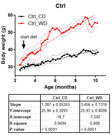
Figure 1: Effect of Western-type diet on body weights of nontransgenic animals.
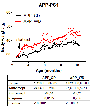
Figure 2: Effect of Western-type diet on body weights of transgenic APP/PS1 animals.
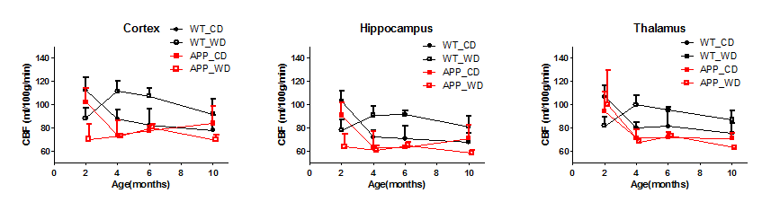
Figure 3: Basal perfusion in the cortex, hippocampus and thalamus of APP-PS1 animals and age-matched controls, divided into Western-type and control diet groups.
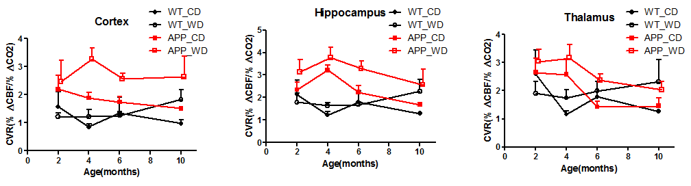
Figure 4: Cerebrovascular response (CVR) to hypoventilation-induced hypercapnia in the cortex, hippocampus and thalamus of APP/PS1 animals and age-matched controls, divided into Western-type and control diet groups.