4114
CEREBELLAR TRACTOGRAPHY STUDY ON ADOLESCENT IDIOPATHIC SCOLIOSIS1Department of Imaging and Interventional Radiology, The Chinese University of Hong Kong, Hong Kong, Hong Kong, 2Department of Medicine and Therapeutics, The Chinese University of Hong Kong, 3Department of Orthopedics and Traumatology, The Chinese University of Hong Kong, 4Department of Imaging and Interventional Radiology, The Chinese University of Hong Kong
Synopsis
Adolescent Idiopathic Scoliosis (AIS) is a 3-dimensional spinal deformity which occurs predominantly in adolescent girls. Impaired postural balance and tonsillar ectopia are reported findings in AIS. This study sought to investigate and compare white matter integrity of the cerebellar pathways in AIS with matched controls. Anatomically-guided deterministic tractography was used to reconstruct the cerebellar pathways. Lower FA value was found in spinocerebellar tract and in intracerebellar tract in AIS, suggestive of, reduced white matter integrity of the cerebellar pathway, which is in agreement with similar changes in other white matter pathways within the brain of AIS patients.
Introduction
Adolescent Idiopathic Scoliosis (AIS) is a 3-dimensional spinal deformity which occurs predominantly in adolescent girls. Despite advances of the surgical treatment for AIS, its etiopathogenesis remains unanswered. While recent studies support AIS is a multifactorial disease, tonsillar ectopia has been found to be common associated feature in AIS patients. Cerebellum is a part of the brain which plays an important role in motor controls, postural balance, cognitive function, language, and somatosensory functions. Moreover, some of the neurophysiologic function correlated to cerebellum dysfunction has been found to be abnormal in AIS, such as the balance control, and somatosensory function, hence, suggesting AIS is probably related to cerebellum dysfunction. Although volumetric change of the cerebellum has been reported in AIS, very little is known about its white matter integrity. The objective of this study is to investigate and understand the cerebellar white matter pathways integrity in AIS using deterministic tractography analysis.Methods
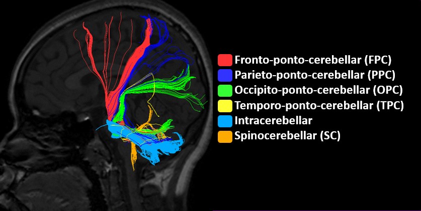
Sixty nine AIS patients (female, right thoracic curve (65), right thoracolumbar curve (4), mean age 14.5 +/- 2.2) and 40 age-matched control subjects (female, mean age 14.6 +/- 1.04) underwent DTI along 32 non-linear directions using a 3T scanner (Achieva TX; Philips Healthcare, Best, The Netherlands). All subjects were right-handed. DTI data were then went through preprocessing and analyzed by using anatomically guided deterministic tractography methods available in DSI Studio[1] to reconstruct spinocerebellar tract (SC), intracerebellar tract and cortico-ponto-cerebellar tract (CPC), where CPC includes the fronto-ponto-cerebellar tract (FPC), occipito-ponto-cerebellar tract (OPC), parieto-ponto-cerebellar tract (PPC), and temporo-ponto-cerebellar tract (TPC). The tracts are shown in figure 1. The Fractional Anisotropy (FA) threshold was set to 0.15, and an angle threshold was set to 700[2]. Regions of interest (ROI) for SC were placed in inferior cerebellar peduncle (ICP) and ipsilateral cerebellar hemispheres, while ROI for intracerebellar tract were placed in both hemispheres of the cerebellum. ROI placement for CPC were in middle cerebellar peduncle (MCP) and each lobe depending on the reconstructed tract, such as for FPC, the other ROI was placed in the contralateral frontal lobe, while PPC used the ipsilateral parietal lobe, OPC used ipsilateral occipital lobe, and TPC used ipsilateral temporal lobe as second ROI. Other than cerebellum where its ROI was manually drawn, other ROIs were using registered MNI atlas and registered JHU White matter atlas. FA value of each tracts and each group were extracted, and compared by using independent t-test. Threshold for the statistical significance was set at p-value = 0.05.Results
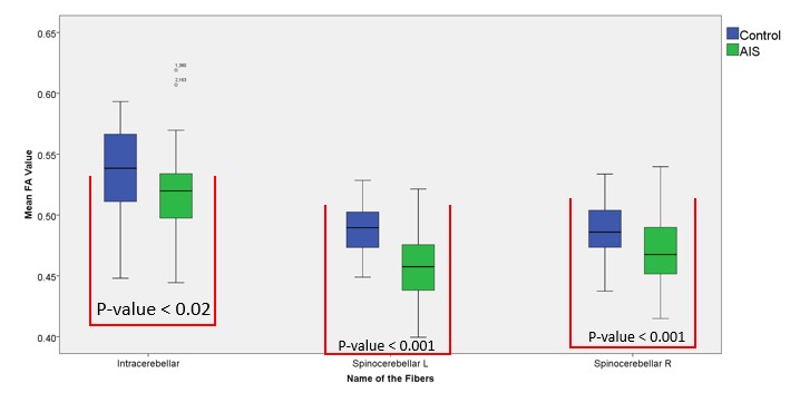
Fiber tractography analysis revealed significantly lower FA values in spinocerebellar pathways (p-value < 0.001) which is projected from the spinal cord to cerebellum and the intracerebellar pathways (p-value < 0.02) interconnecting left and right hemispheres of the cerebellum. The FA value of the all cortico-ponto-cerebellar pathways was not found significantly different when comparing to control subjects.Discussion
Morphological changes in cerebellum and low lying cerebellar tonsils [3, 4] have previously been reported in AIS. In this study, we found lower FA value in spinocerebellar tract as well as intracerebellar tract in AIS comparing to the control subjects. Since spinocerebellar tract plays a role in relaying proprioception and sensorimotor information[5], lower FA value in spinocerebellar tract might correlate to alteration of sensorimotor integration, while lower FA value found in the intracerebellar pathway indicates reduction of white matter integrity/ connectivity between both sides of the cerebellum. Both the above changes might reflect some form of cerebellar dysfunction and contribute to impaired postural balance in AIS [6]. Furthermore, the lower FA values in spinocerebellar tract coincides with the finding of reduced FA value in medulla oblongata and cervical spinal cord in AIS patients when compared with controls, as previously reported by Kong, et.al[7], in which lower tonsillar level was also consistently found.Conclusion
Reduced white matter integrity with lower FA values was found in spinocerebellar tract and intracerebellar tract in AIS, which might be related to abnormal postural balance control in this group of patients.Acknowledgements
This research is supported by SHHO Scoliosis Research Laboratory (Award No. 7104486), and a grant from Kai Cheong TongReferences
[1] F. C. Yeh, T. D. Verstynen, Y. Wang, J. C. Fernandez-Miranda, and W. Y. Tseng, "Deterministic diffusion fiber tracking improved by quantitative anisotropy," PLoS One, vol. 8, p. e80713, 2013.
[2] Z. Keser, K. M. Hasan, B. I. Mwangi, A. Kamali, F. E. Ucisik-Keser, R. F. Riascos, N. Yozbatiran, G. E. Francisco, and P. A. Narayana, "Diffusion tensor imaging of the human cerebellar pathways and their interplay with cerebral macrostructure," Front Neuroanat, vol. 9, p. 41, 2015.
[3] L. Shi, D. Wang, S. C. Hui, M. C. Tong, J. C. Cheng, and W. C. Chu, "Volumetric changes in cerebellar regions in adolescent idiopathic scoliosis compared with healthy controls," Spine J, vol. 13, pp. 1904-11, Dec 2013.
[4] W. C. Chu, G. C. Man, W. W. Lam, B. H. Yeung, W. W. Chau, B. K. Ng, T. P. Lam, K. M. Lee, and J. C. Cheng, "A detailed morphologic and functional magnetic resonance imaging study of the craniocervical junction in adolescent idiopathic scoliosis," Spine (Phila Pa 1976), vol. 32, pp. 1667-74, Jul 1 2007.
[5] B. L. Riemann and S. M. Lephart, "The sensorimotor system, part I: the physiologic basis of functional joint stability," J Athl Train, vol. 37, pp. 71-9, Jan 2002.
[6] M. Simoneau, N. Richer, P. Mercier, P. Allard, and N. Teasdale, "Sensory deprivation and balance control in idiopathic scoliosis adolescent," Exp Brain Res, vol. 170, pp. 576-82, Apr 2006.
[7] Y. Kong, L. Shi, S. C. Hui, D. Wang, M. Deng, W. C. Chu, and J. C. Cheng, "Variation in anisotropy and diffusivity along the medulla oblongata and the whole spinal cord in adolescent idiopathic scoliosis: a pilot study using diffusion tensor imaging," AJNR Am J Neuroradiol, vol. 35, pp. 1621-7, Aug 2014.
Figures