4112
VOLUMETRIC BRAIN FINDINGS IN CHILDREN WITH SEIZURES1Diagnostic and Interventional Imaging, KK Women's and Children's Hospital, Singapore, Singapore
Synopsis
Retrospective study of MRI brains with volumetric measurements done using voxel based morphology was conducted for children with seizures imaged in KK Women’s and Children’s Hospital since 2006. There was decreased cerebral volume and increased ventricular volume in children with hippocampal abnormalities compared to children with structurally normal brain, the difference in volumes exceeding 1 standard deviation, not statistically significant likely on account of the small sample sizes. Relative reduced grey matter to white matter is seen in children with hippocampal abnormalities as well as in children with neuronal migration abnormalities, reaching statistical significance in both groups.
Purpose
To quantify the volumetric changes seen in MRI of children with seizures with voxel based morphology.Methods
This retrospective study was approved by the institutional review board with waiver of consent obtained. Paediatric patients who had volumetric high-resolution anatomical T1 weighted imaging on either a 3T MR scanner (MAGNETOM Skyra, Siemens Healthcare) or on a 1.5T MR scanner (General Electric Signa Excite Mallinkrodt ) since 2006 were selected for analysis. Infants were excluded as the myelination process is still in progress on T1 weighted images. All anatomical images were post processed by voxel based morphometry to generate volumes of ventricles, cerebral parenchyma, grey matter and white matter. The percentages of contributed by ventricles, cerebral parenchyma, grey matter and white matter and hippocampi were compared between those with structurally normal brains and those with abnormalities as defined in the MRI report. Statistical analysis was performed on IBM SPSS Statistics version 19 using 2 tailed T test with unequal variance with significance level set as p≤0.05.Results
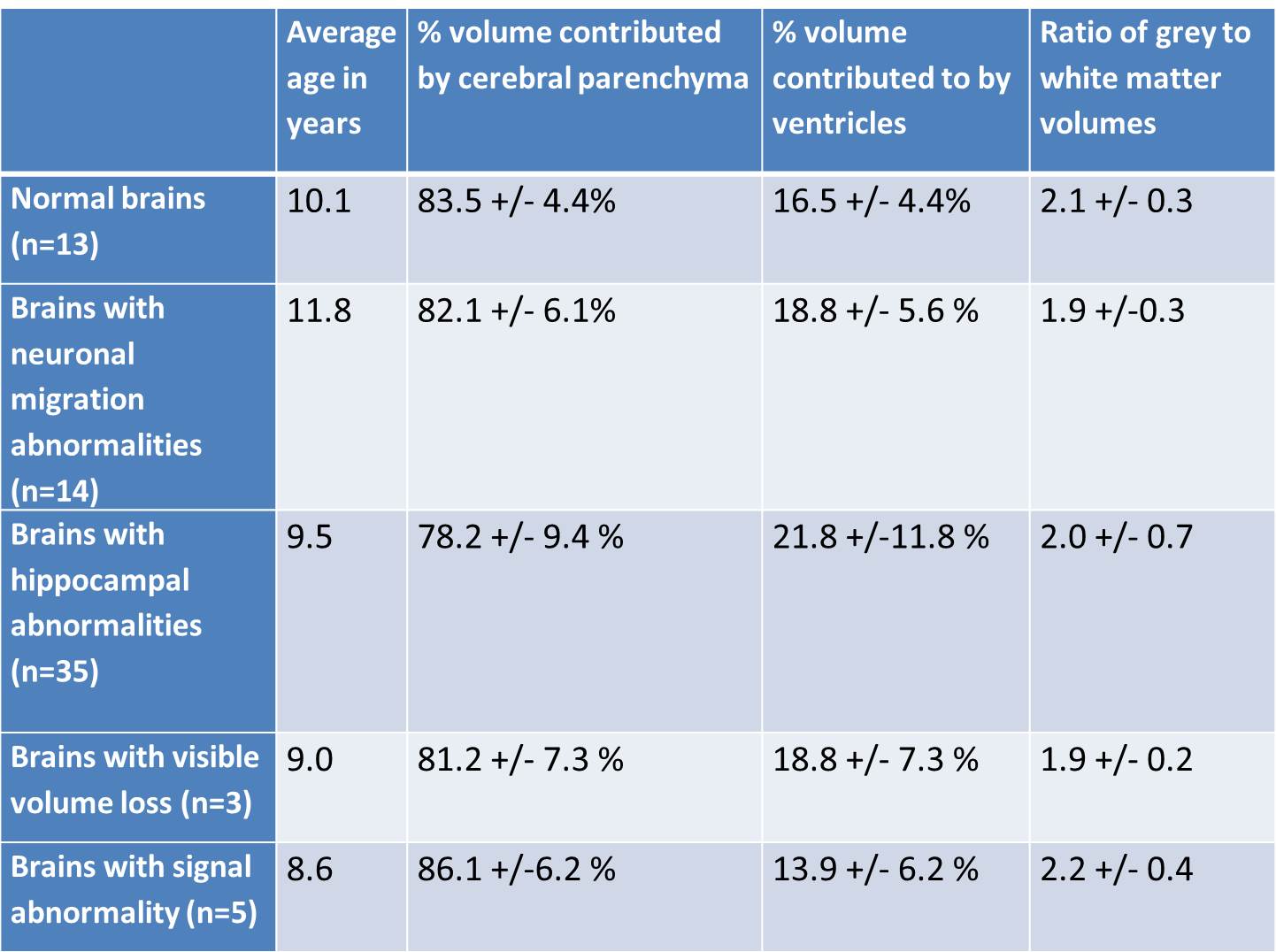
Seventy paediatric patients aged 1 year old to 21 years old ( average 10 years, 31/70 males) had MRI done either on a 3T MR scanner (MAGNETOM Skyra, Siemens Healthcare) with a 32-channel head coil or on a 1.5T MR scanner (General Electric Signa Excite Mallinkrodt ) with a 8 channel head coil. All had high-resolution anatomical volumetric T1 weighted images. Thirteen patients had structurally normal brains based on MRI report. Fifty seven patients had abnormalities on the scans, some having more than 1 type of abnormality. There were 35 with hippocampal abnormality (6 right side, 18 left sided, 11 bilateral), 14 with neuronal migration abnormality (2 on right side, 6 left sided, 6 bilateral), 3 with volume loss, 5 with signal abnormality. For paediatric seizure patients with normal brains on MRI, cerebral parenchyma accounted for 83.5 +/- 4.4% and ventricles 16.5 +/- 4.4% and the average grey to white matter ratio was 2.1 +/- 0.3. Volumes and ratio of grey to white matter volumes with respect to visible MRI abnormalities are presented in Table 1. There was statistically significant differences present in the following Children with bilateral hippocampal abnormalities showed lower grey to white matter ratio of 1.7 compared to those with structurally normal brains which had ratio of 2.1 (p=0.02) Children with left hippocampal signal abnormality showed lower grey to white matter ratio of 1.7 compared to those with structurally normal brains which had ratio of 2.1 (p=0.03) Children with right neuronal migration abnormalities showed lower grey to white matter ratio of 1.9 compared to those with structurally normal brains which had ratio of 2.1 (p=0.04).Discussion
MRI is the modality of choice for seizure imaging and most common abnormalities described in the literature are ventricular enlargement, gliosis, heterotopias and cortical dysplasia.1 Ratios were used to compare across the various ages in our study population as actual volumes of grey and white matter are known to change with brain development in children.2 Although there was decreased cerebral volume and increased ventricular volume in children with hippocampal abnormalities compared to children with structurally normal brain and the difference in volumes exceeding 1 standard deviation, the difference was not statistically significant likely on account of the small sample sizes. Limitation of this study is the lack of a control population of healthy children for direct comparison with the patient population and the small sample sizes in each of our subgroups. Statistical significance reduced grey matter to white matter ratio is seen in children with hippocampal abnormalities as well as in children with neuronal migration abnormalities. A control population would allow finer analysis as to whether this is due to decreased grey matter or increased white matter volumes.Conclusion
Quantifiable decreased cerebral volume and increased ventricular volume in children with hippocampal abnormalities compared to children with structurally normal brain.Acknowledgements
Many thanks to students Ng ML and Chan YM for their help in the volumetric measurements.References
1. Kalnin A, Fastenau PS, deGrauw TJ, et al. MR Imaging Findings in Children with First Recognized Seizure. Pediatr Neurol. 2008;39(6):404–414.
2) Herman BP, Dabbs K, Becker T et al. Brain development in children with new onset epilepsy: A prospective controlled cohort investigation Epilepsia. 2010;51(10):2038–2046.
Figures