4107
The type and prevalence of incidental findings on magnetic resonance imaging of the low risk term born neonatal brain.1Perinatal Imaging and Health, Kings College London, London, United Kingdom, 2Paediatric radiology, Great Ormond Street Hospital, London, United Kingdom, 3Biomedical engineering and imaging sciences, Kings College London, London, United Kingdom
Synopsis
The aim of this study was to ascertain the prevalence and type of incidental findings present in the low risk term brain using MRI. Out of 200 infants, 105 had incidental findings, with 8 requiring follow-up assessment. Common findings included subdural haemorrhage, punctate and cystic lesions and hyperintense signal of the basal ganglia. Findings requiring further assessment included subarachnoid, cerebellar, and germinal matrix haemorrhages, ectopic posterior pituitary lobe, subepenymal heterotopia and venous infarcts. Prevalence of incidental findings in this cohort is significant. Communicating to parents the possibility of detecting abnormalities and referral pathways are important considerations in neonatal research.
Purpose
Asymptomatic intracranial abnormalities of potential clinical or non-clinical significance are relatively common in many adult and paediatric magnetic resonance imaging (MRI) research studies of the brain(1) . The overall prevalence of incidental findings in both adult and paediatric populations is approximately 20% (1,2), with findings that require further investigation or a neurological referral reported at 2-3%(3). However, within the paediatric studies, the prevalence and type of incidental findings that are present in the ‘low risk’ term born infant using 3T high resolution MRI have not yet been determined. The developing Human Connectome Project (dHCP) aims to collect up to 1500 high-resolution structural brain scans using MRI from fetuses and newborn infants, including 500 healthy term infants. The aim of this study was to ascertain from this cohort, the prevalence and type of incidental findings present in the 'low risk' term born brain.Methods
Participants
From the 6th of February 2015 until the 13th of June 2016, 366 infants underwent brain MRI as part of the dHCP. From this cohort, the first 200 term (gestational age (GA) >36+6 weeks) low risk infants were included in the analysis. Clinical characteristics of the study group: GA range at birth: (37 weeks-42+2 weeks); age range at scan: (37+2 weeks-44+6 weeks), gender: 112 (56%) male, mode of delivery: n=70 (35%) spontaneous vaginal, n=25(12.5%) elective cesarean section, n=64(32%) emergency cesarean section, n=28(14%) forceps, and n=13(6.5%) ventouse.
Imaging
All 200 infants underwent MRI imaging in natural sleep using a dedicated neonatal 32-channel phased array head coil(4) on a Philips 3T Achieva system. Structural imaging consisted of an axial and sagittal T2 TSE (turbo spin echo) sequence (TR/TE=12000/156ms, acquired voxel size = 0.8X0.8x1.6mm, slice gap = -0.8mm, TSE factor =12, number of slices =125 (axial and 145 sagittal), profile order=asymmetric, SENSE factor= 2.1, time =3.12minutes) and a sagittal 3D MPRAGE (TR/TE/TI= 11/4.6/713ms, flip angle=90, acquired voxel size =0.8x0.8x0.8mm, number of slices =135, SENSE factor=1.2, time=4.35mins). All imaging raw data was reconstructed at the time of acquisition using the standard scanner software and then also using motion correcting software(5).
Image analysis
A random sample of 40, T2 axial and sagittal images were graded for motion artefacts before and after motion correction. Both the non motion-corrected and motion-corrected images were reported by an experienced neonatal radiologist. Where incidental findings were suspected the motion-corrected images were used as an additional diagnostic tool. Abnormal appearances to the brain were noted and discussed with a multidisciplinary team to decide on appropriate additional investigations and follow-up.
Results
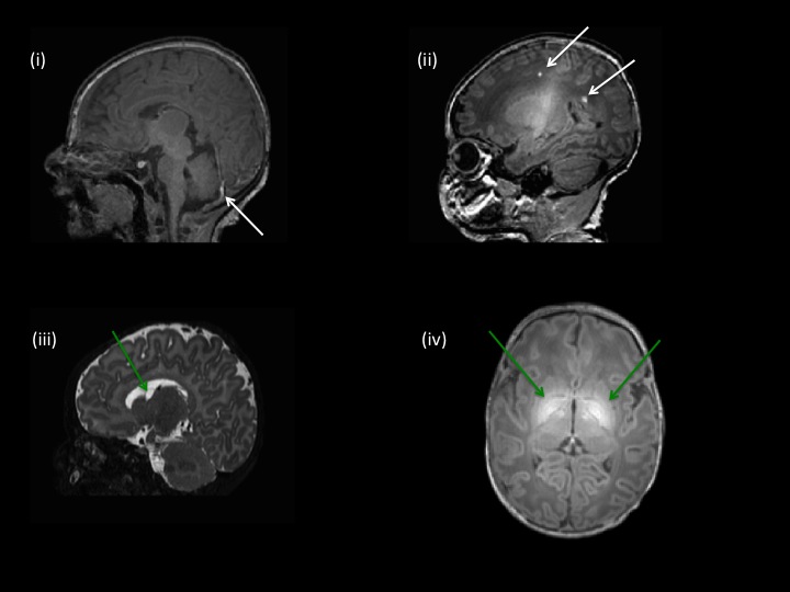
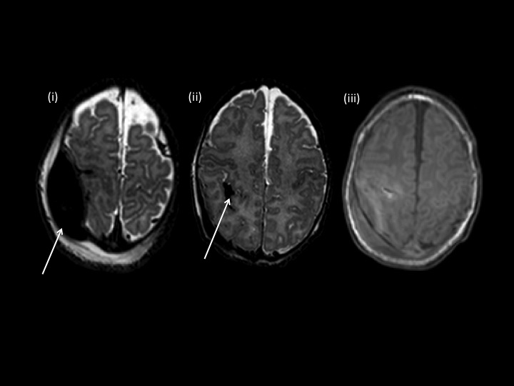
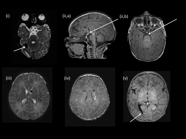
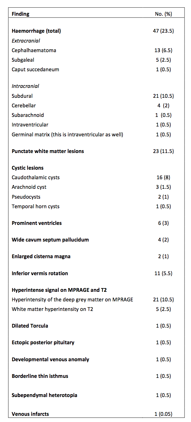
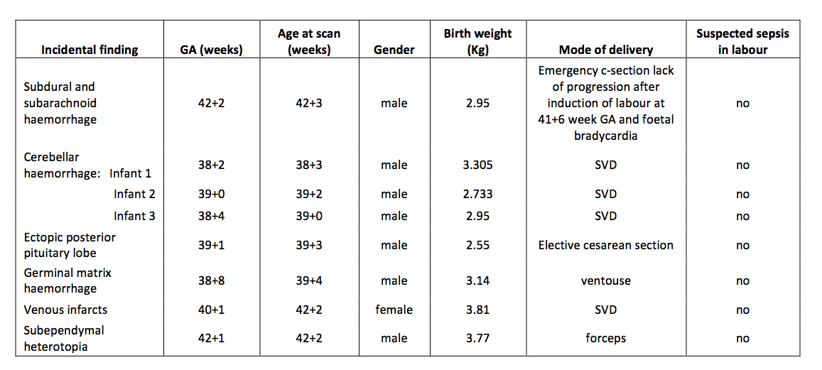
Out of the 200 low risk term born infants, 105 (52.5%) were reported as having one or more incidental finding on their structural images, with 8 (4%) of the infants requiring repeat imaging and/or follow-up by neonatal/neurology consultant. The type and prevalence of the incidental findings can be found in Table 1. MR imaging examples of the most common findings that included subdural haemorrhage, punctate and cystic lesions and hyperintense signal of the globi pallidi and subthalamic nuclei on MPRAGE images are shown in Figure 1. Incidental findings that required referral to a neonatal/neurology consultant included subarachnoid, cerebellar, and germinal matrix haemorrhages, an ectopic posterior pituitary lobe, subepenymal heterotopia and venous infarcts. MR imaging examples of these findings are shown in Figures 2 and 3. Clinical characteristics of each infant that required repeat imaging and/or follow-up can be found in table 2. Motion artefact assessment: 60% of scanner T2 axial and 80% of T2 sagittal had motion present. In all but 1 case, motion correction completely resolved the artefacts. The impact of this for radiological assessment has not been assessed as yet.Conclusions
Incidental findings in the low risk term born infant brain using MRI is higher than that reported in adults and older paediatric populations. In this study, 52.5% of infants had reported incidental findings with 4% requiring further investigation. Self-selection on the part of the volunteering parents may have skewed the sample relative to a simple cross section of deliveries and this is something that will be further investigated. Likewise, the impact on radiological findings of motion artefact, which was common in the scanner reconstructions for our unsedated neonates and the benefit of correcting this in reconstruction remains to be formally assessed. Nevertheless, these results highlight the importance of communicating to parents beforehand the possibility of detecting an incidental finding on their newborn infants’ research MRI scan and ensuring that adequate referral pathways are in place.Acknowledgements
No acknowledgement found.References
(1) Incidental findings on brain magnetic resonance imaging: systematic review and meta-analysis
Zoe Morris, William N Whiteley, W T Longstreth Jr, Frank Weber, Yi-Chung Lee, Yoshito Tsushima, Hannah Alphs, Susanne C Ladd, Charles Warlow, Joanna M Wardlaw, Rustam Al-Shahi Salman. BMJ 2009;339:b3016
(2) Incidental findings are frequent in young healthy individuals undergoing magnetic resonance imaging in brain research imaging studies: a prospective single-center study.
Hartwigsen G1, Siebner HR, Deuschl G, Jansen O, Ulmer S. J comput Assit Tomogr. 2010 Jul;34(4):596-600.
(3) Incidental Findings on Pediatric MR Images of the Brain Brian.
S. Kim, Judy Illes, Richard T. Kaplan, Allan Reiss, and Scott W. Atlas AJNR Am J neuroradiol 23:1674-1677, 2002.
(4) A dedicated neonatal brain imaging system
Hughes, E. J., Winchman, T., Padormo, F., Teixeira, R., Wurie, J., Sharma, M., Fox, M., Hutter, J., Cordero-Grande, L., Price, A. N., Allsop, J., Bueno-Conde, J., Tusor, N., Arichi, T., Edwards, A. D., Rutherford, M. A., Counsell, S. J. and Hajnal, J. V. (2016), Magn. Reson. Med.. doi:10.1002/mrm.26462.
(5) Sensitivity encoding for aligned multishot multislice MRI
Cordero-Grande L, Teixera, RPAG, Hughes EJ, Hutter J, Price AN, Hajnal JV. Sensitivity encoding for aligned multishot multislice MRI 2016; IEEE Transactions on Computational Imaging.
Figures