4106
Neonatal whole brain volume and microstructure in infants born preterm and neurodevelopmental outcomes at 2 years of age1Murdoch Childrens Research Institute, Melbourne, Australia, 2Newborn research, Royal Women’s Hospital, Melbourne, Australia, 3Department of Obstetrics and Gynaecology, The University of Melbourne, Melbourne, Australia, 4Department of Physiotherapy, The University of Melbourne, Melbourne, Australia, 5Department of Medicine, Monash Medical Centre, Monash Univeristy, Melbourne, Australia, 6Department of Paediatrics, The University of Melbourne, Melbourne, Australia, 7Florey Institute of Neuroscience and Mental Health, Melbourne, Australia
Synopsis
Infants born preterm are at risk of neurodevelopmental delays in childhood, and MRI may improve knowledge of underlying cerebral changes. Relationships were investigated between whole brain volumes and microstructure in 256 preterm infants and neurodevelopmental outcomes at age 2 years using voxel-based morphometry and tract-based spatial statistics. Lower grey and white matter volumes and altered white matter microstructure were associated with poorer outcomes. In general, relatively widespread brain regions were associated with cognition, more central regions with cerebral palsy, and more peripheral regions with language. This study provides further understanding of how brain structure in preterm infants is related to longer-term outcomes.
Introduction
Infants born preterm [<37 weeks’ gestational age (GA)] are at high risk of having problems in childhood in wide-ranging neurodevelopmental domains, including cognition, movement and behaviour.1 Many previous studies have focused on very preterm infants (VPT, <32 weeks’ GA), however recent research suggests that moderate and late preterm infants (MLPT, 32-36 weeks’ GA) are also at increased risk of long-term neurodevelopmental problems compared with full-term infants.2 Defining brain structure – function relationships in preterm infants, including VPT and MLPT infants, may improve knowledge of the factors contributing to neurodevelopmental outcomes and the infants who are most likely to have poorer outcomes. The aim of the current study was to investigate whether whole brain structure and microstructure in preterm infants is associated with cerebral palsy and neurodevelopmental outcomes at 2 years of age, and whether these associations differ for VPT and MLPT infants.Methods
150 VPT (<30 weeks’ GA) and 201 MLPT (32-36 weeks’ GA) infants were recruited from the Royal Women’s Hospital, Melbourne, into prospective cohort studies. Of those recruited, 104 VPT and 198 MLPT infants underwent magnetic resonance imaging (MRI) between 38-44 weeks’ GA inclusive. The participants’ neurodevelopment was assessed at 2 years of age (corrected for prematurity); cerebral palsy was diagnosed by paediatricians, and the Bayley Scales of Infant and Toddler Development was administered to assess cognitive, language and motor development. The current study included 251 infants (88 VPT and 163 MLPT) with both neonatal structural images and 2-year neurodevelopmental data, and 256 infants (85 VPT and 171 MLPT) with both neonatal diffusion images and 2-year neurodevelopmental data. Structural brain images were segmented into tissue types using the Morphologically Adaptive Neonatal Tissue Segmentation (MANTiS) technique.3 Cortical grey matter and white matter maps in neonatal template space4 were analysed using Voxel-Based Morphometry (VBM). Diffusion images were corrected for echo planar imaging and motion/eddy current distortions, and the diffusion tensor model was fitted. Diffusion tensor images [fractional anisotropy (FA) and axial (AD), radial (RD) and mean (MD) diffusivities] were analysed using Tract-Based Spatial Statistics,5 where all images were registered to the most representative image in the cohort. Whole-brain, voxel-wise statistical analysis of volumes and diffusion measures was performed using non-parametric permutation based methods.6 Associations between neonatal volume or diffusion measures and 2-year outcomes were analysed, adjusted for the potential confounder of age at MRI, as well as sex for volumes. Volume analyses were also performed with and without adjusting for intracranial volume (ICV). Additionally, differences in brain structure-function relationships between VPT and MLPT infants were investigated by interaction analyses. Results are reported at p<0.05 following 5000 permutations, threshold-free cluster enhancement and family-wise error rate correction. Regions of statistical significance were localised to anatomical brain regions and tracts by visual inspection and comparison against a neonatal atlas.7Results
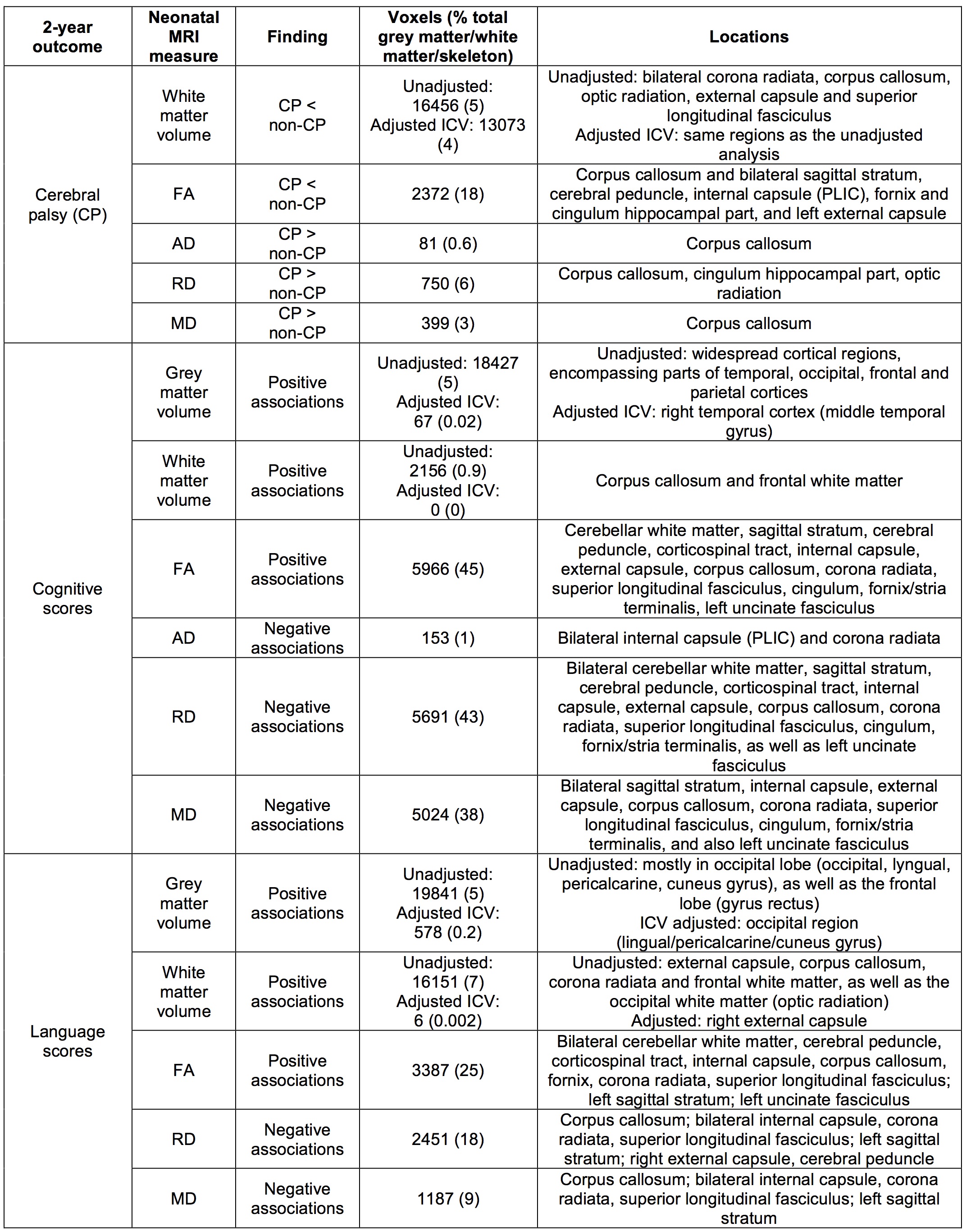
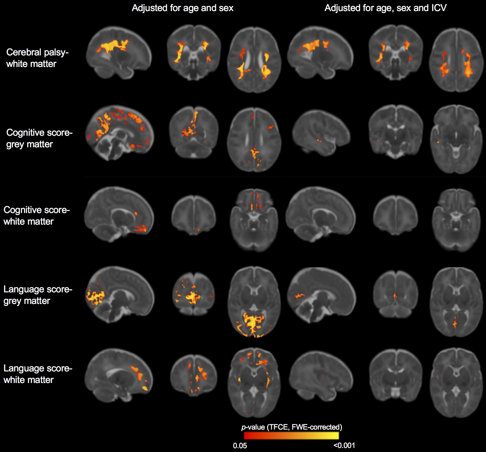
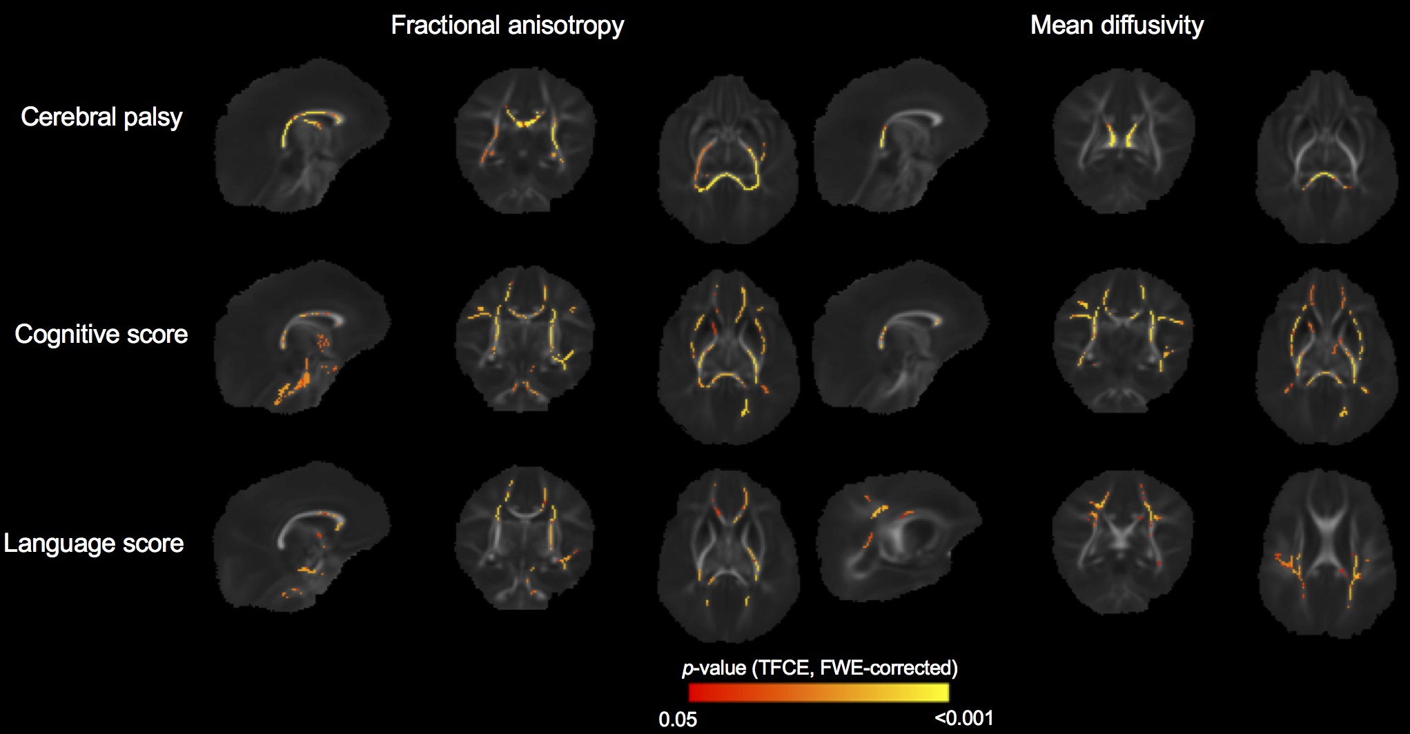
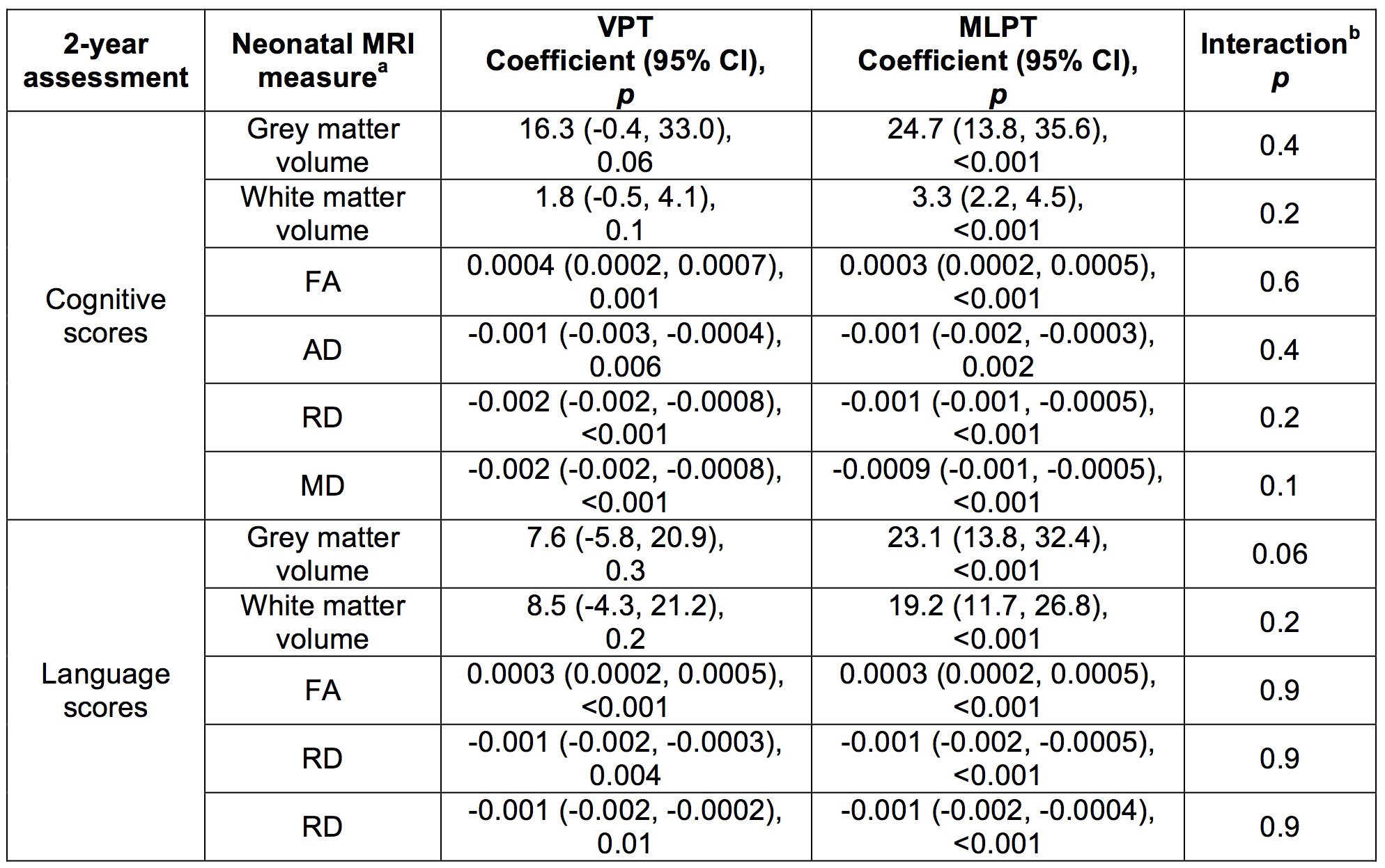
In children who had cerebral palsy compared with children who did not, white matter volume and FA were lower, and AD, RD and MD were higher, in many regions listed in Table 1 and shown in Figures 1 and 2. Higher grey matter volume, white matter volume and FA, and lower AD, RD and MD, in preterm infants were associated with higher (better) cognitive and language scores at 2 years of age (Table 1, Figures 1 and 2). In general, relatively widespread brain regions were associated with cognitive scores, while more central and motor regions (e.g. corpus callosum, corona radiata) were associated with cerebral palsy, and more peripheral regions (e.g. superior longitudinal fasciculus and frontal and occipital cortices) were associated with language outcomes (Table 1, Figures 1 and 2). There was little evidence of associations between MRI measures in preterm infants and motor development at 2 years of age. For volumes, many findings remained after adjusting for ICV, although some findings weakened (Table 1, Figure 1). In general, the associations between brain measures and neurodevelopmental outcomes were similar between the VPT and MLPT groups, although the relationships with volumes were stronger in the MLPT than VPT infants (Table 2).Discussion
MRI measures of whole brain structure and microstructure at term-equivalent age in both VPT and MLPT infants are associated with neurodevelopmental outcomes two years later. Associations were identified in widespread brain regions and white matter tracts, which differed depending on the functional outcome.Conclusion
This study provides greater knowledge of brain structure-function relationships in children born preterm. Such knowledge may aid early identification of infants who are at greatest risk for developing problems later in life.Acknowledgements
No acknowledgement found.References
1. Saigal S and Doyle LW. An overview of mortality and sequelae of preterm birth from infancy to adulthood. Lancet. 2008;371(9608):261-269.
2. Cheong JLY, Doyle LW. Increasing rates of prematurity and epidemiology of late preterm birth. J Paediatr Child Health. 2012;48(9):784-788.
3. Beare RJ, Chen J, Kelly CE, et al. Neonatal Brain Tissue Classification with Morphological Adaptation and Unified Segmentation. Front Neuroinform. 2016;10(12). doi: 10.3389/fninf.2016.00012.
4. Kuklisova-Murgasova M, Aljabar P, Srinivasan L, et al. A dynamic 4D probabilistic atlas of the developing brain. NeuroImage. 2011;54(4):2750-2763.
5. Smith SM, Jenkinson M, Johansen-Berg H, et al. Tract-based spatial statistics: voxelwise analysis of multi-subject diffusion data. NeuroImage. 2006;31(4):1487-1505.
6. Winkler AM, Ridgway GR, Webster MA, et al. Permutation inference for the general linear model. NeuroImage. 2014;92:381-397.
7. Oishi K, Mori S, Donohue PK, et al. Multi-contrast human neonatal brain atlas: application to normal neonate development analysis. NeuroImage. 2011;56(1):8-20.
Figures