4103
Music training enhances functional connectivity in preterm newborns1Division of Development and Growth, Department of Pediatrics, University Hospital of Geneva, Geneva, Switzerland, 2Institute of Bioengineering, Ecole Polytechnique Fédérale de Lausanne, Lausanne, Switzerland, 3Neuroscience of Emotion and Affective Dynamics Lab, Department of psychology, University of Geneva, Geneva, Switzerland, 4Swiss center for affective neurosciences, University of Geneva, Geneva, Switzerland, 5Department of Radiology and Medical Informatics, University of Geneva, Geneva, Switzerland
Synopsis
In this study we examined the impact of the environmental enrichment of preterm newborns with music on auditory cortex functional connectivity. A group of preterm infants listened to music from 33 weeks gestational age until term equivalent age. Two control groups were used: preterm and full-term infants without music. Auditory cortex functional connectivity with cerebral regions known to be implicated in tempo and familiarity processing were identified for preterm newborns that had music training during their stay in the NICU using Psychophysiological Interaction (PPI) analyses. Our results suggest that music training during hospitalization can modify functional connectivity in the newborn brain.
Purpose
Neural and functional consequences of prematurity warrant for consistent neonatal intensive care unit(NICU) enhancement. Appropriate environmental enrichment has been shown to have positive impact on behavioral states and brain development1. Over the past decade, music in NICU has been shown to positively impact on physiological and behavioral measures2. Furthermore, fetal auditory learning has been demonstrated in newborns when listening to the same music they heard during last trimester of gestation but with some notes changed3. Thus, the aim of this study is to observe the impact of an early environmental enrichment of preterm infants with music on auditory cortex functional connectivity and to assess if preterm infants can already learn to process subtle changes in this music like tempo modification.Methods
Subjects:Three groups of newborns were recruited. Music group consisted of 18preterm newborns (mean GA: 28.83wks±2.14) which were listening to music with headphones five times per week during the hospitalization in the NICU. Non-music group consisted of 17preterm newborns (mean GA: 29.11wks±1.85) which had headphones without music. Term group consisted of 21full-term newborns (mean GA: 39.44wks±1.12).
Acquisition:MRI scan was conducted at birth for full-term infants and term-equivalent-age for preterm groups, without any sedation. Siemens 3T scanners (Siemens Trio; Siemens Prisma) were used to obtain T2*-weighted gradient-echo EPI images (TR=1600ms, TE=30ms, 30slices, voxel-size=2.5x2.5x3.0mm3) as well as T2-weighted TSE image for anatomical reference (TR=4600ms, TE=150ms, 113coronal slices, voxel-size=0.78x0.78x1.2mm3). Music stimuli were presented 10 times in a pseudo-random block design protocol during 8seconds (5conditions: Silence, Background-Music, Original-Music, Transposed-Music (key modulation), and Tempo-Modification (Original-Music played 40%faster)).
Prepossessing:Realignment, slice-timing, coregistration, normalization and smoothing(6-mm FWHM). 24motion-related parameters (6realignment parameters+Volterra expansion) were also included in the model as covariates to remove any residual motion-related variance4. Only infants with absolute motion <1mm for more than 70%of images were used for subsequent analyses. Infants were excluded because of major brain lesions. Finally, all subsequent analyses were performed only on infants showing activations induced by the overall sound-versus-silence contrast, at p<0.01 uncorrected in auditory regions, resulting in 9 infants for each group.
Analyses:Psychophysiological Interaction(PPI) analyses were conducted. The experimental conditions, the baseline and the motion regressors were included in the design matrix. After defining the contrasts of interest (e.g. Original-Music vs Tempo-Modification), BOLD signals were extracted from Left(L_A1) and Right(R_A1) Primary Auditory cortex for each subject. Then PPI analyses were carried out for each subject using the gPPI toolbox which also supports standard PPI analysis5. Finally, subject-specific contrast images using the contrast(0-1-0), where the second column in the design matrix represented the PPI term, were then entered into second level analyses (corrected for multiple comparisons at cluster level with a threshold of p<0.005uncorrected and extent-threshold 20voxels).
Results
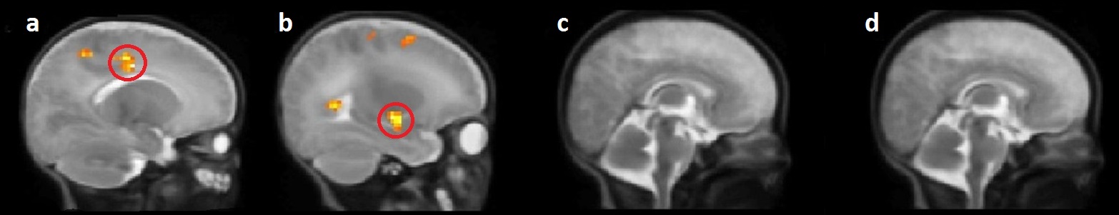
One-sample t-tests: For the Original music vs Tempo modification contrast and the L_A1 as seed region, we found statistically significant activation in the left middle-cingulate-cortex(MCC) and in the right caudate nucleus and putamen for the Music group. No statistically significant results were found for the Non-music and the Term group for both seeds(Fig.1).
Two-sample t-tests:
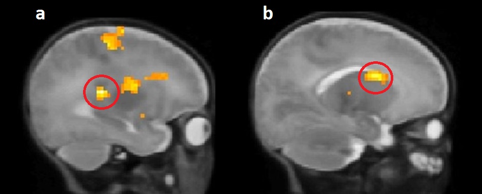
Music>Non music group: Seed R_A1: we found statistically significant activation in the right thalamus, left-MCC and left caudate nucleus(Fig.2).
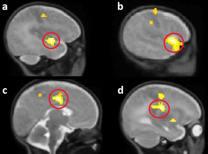
Music>Term group: Seed L_A1: we found statistically significant activations in the superior temporal gyrus(STG) and in the left-MCC. Seed R_A1: statistically significant activations in the left-MCC and putamen (Fig.3).
Note that no statistically significant results were found for the Non-music>Term, Term>Non music , Non-music>Music, Term>Music.
Discussion and Conclusions
Functional connectivity differences when listening to Original-Music compared to Tempo-Modification where found only in the Music group and not in full-term or preterm infants without music exposure, indicating that these differences in music processing may rely on learning due to musical training rather than innate tempo detection ability.
Thalamus and basal ganglia have been shown to be implicated in tempo processing in adults. Additionally, putamen has been shown to increase its functional connectivity with STG when listening to beat stimuli compared to non-beat stimuli6. Also, activation of MCC and STG for familiar music has been shown to be dependent on musical expertise7.
Taken together, these results indicate that enrichment of the NICU with music can have long lasting learning effects on music processing with an increased connectivity between primary auditory cortex and brain regions implicated in music tempo and familiarity processing.
To our knowledge, this study is the first one using psychophysiological interaction (PPI) analysis on newborns and showing that preterm newborns can learn tempo discrimination by listening to music during their stay in neonatology using fMRI. Our results indicate that music enrichment of the NICU’s environment can modify functional connectivity in the newborn brain.
Acknowledgements
No acknowledgement found.References
1. Als H, Duffy FH, McAnulty GB, et al. Early experience alters brain function and structure. Pediatrics.2004;113(4):846-857.
2. Halsbeck FB. Music therapy for premature infants and their parents: an integrative review. Nordic Journal of Music Therapy. 2012; 21(3):203-226.
3. Partanen E, Kujala T, Tervaniemi M, et al. Prenatal Music Exposure Induces Long-Term Neural Effects. PLoS ONE.2013;8(10): e78946
4. Friston KJ, Williams S, et al. Movement-related effects in fMRI time-series. Magn Reson Med.1996; 35(3):346-355.
5. McLaren DG, Ries ML, Xu G, et al. A Generalized Form of Context-Dependent Psychophysiological Interactions (gPPI): A Comparison to Standard Approaches. Neuroimage.2012; 61(4):1277-1286.
6. Grahn JA and Rowe JB. Feeling the Beat: Premotor and Striatal Interactions in Musicians and Nonmusicians during Beat Perception. Journal of Neuroscience.2009; 29(23):7540-7548.
7. Groussard M, Joie R, Rauchs G, et al. When Music and Long-Term Memory Interact: Effects of Musical Expertise on Functional and Structural Plasticity in the Hippocampus. PLOS.2010; 5(10):e13225.
Figures