4100
Axon development revealed by directional diffusivities in intra-axonal and extra-axonal compartments1Department of Radiology, the First Affiliated Hospital, Xi'an Jiaotong University, Xi'an, People's Republic of China, 2Department of Biomedical Engineering, the Key Laboratory of Biomedical Information Engineering of the Ministry of Education, School of Life Science and Technology, Xi'an Jiaotong University, Xi'an, People's Republic of China, 3MR Research China, GE Healthcare, Bei Jing, People's Republic of China
Synopsis
White matter in human brain undergoes a complex and long lasting process. It is not likely that conventional diffusion parameters are specific enough to distinguish axon-related and myelin-related processes. This study investigates directional diffusivities in intra-axonal and extra-axonal compartments on preterm neonates, term neonates, infants, and adults. Two change patterns of extra-axonal axial and radial diffusivities are found during development. This may be related to premyelination and myelination. Developmental changes of the fiber dispersion and intra-axonal diffusivities are observed in both pre-myelination and myelination periods. Directional diffusivities in intra-axonal and extra-axonal compartments provide more detailed information to evaluate axonal development.
INTRODUCTION
White matter in human brain undergoes a complex and long lasting developmental process 1, 2. The increase of the axon caliber and the myelin sheath forms the basis for the rapid neural transmission 3. Assessing axonal development is critical for understanding the typical development and its related disorders 1, 4. However, it is not likely that conventional diffusion parameters are specific enough to distinguish axon-related and myelin-related processes 2. Directional diffusivities in intra-axonal and extra-axonal compartments may contain valuable information to overcome the above limitation 5, 6. This study tried to characterize the developmental changes in specific metrics on white matter of neonates, infants, and adults.METHODS
This study was approved by the local Institutional Review Board. Before the MRI scan, parents of neonates and infants were informed about the goal and risks involved in the MR scan. Written informed consents were obtained from adults and parents of the neonates and infants. Subjects who were confirmed or suspected to have congenital malformations of central nervous system, congenital infections, metabolic disorders, abnormal appearances in conventional MRI were excluded.
The MRI datasets used in this study were acquired for the clinical examination and diagnosis. To reduce the head movement and complete the MRI procedure, the neonates and infants patients were sedated with a relatively low dose of oral chloral hydrate (25−50 mg/kg). The patient selection, monitoring, and management were performed following the guidelines. Single-shot EPI diffusion kurtosis imaging was performed on a 3.0T scanner (General Electric Signa HDXT, WI, USA) with an eight-channel head coil. The other parameters were: b values = 500, 1000, 2000, 2500 s/mm2; 18 gradient directions; TR/TE = 8000~11000/91.7~126.1 ms; thickness = 4 mm; FOV = 180 × 180 mm2 ~ 240 × 240 mm2 (according to brain sizes); acquisition matrix = 128 × 128 ~ 172 × 172 (to keep the same resolution). Diffusion and kurtosis tensors were estimated by using constrained weighted linear least squares. The intra-axonal axial diffusivity (ADa), intra-axonal radial diffusivity (RDa), fiber dispersion (FD), extra-axonal axial diffusivity (ADe), and extra-axonal radial diffusivity (RDe) were calculated according to the white matter model for DKI 5, 6.
Three regions of interests (ROIs) were selected by using the white matter labels 7: left corticospinal tract (CST), splenium of corpus callosum (SCC), and left inferior fronto-occipital fasciculus (IFO). Mann-Whitney U Test was used to evaluate differences in GA, postnatal ages, postmenstrual age (PMA), and regional values of metrics across groups. The relative changes in metrics were calculated by: 100% × (values in group 2−values in group 1)/values in group 1. Tests were considered significant at P<0.05.
RESULTS
A total of 17 preterm neonates, 48 term neonates, 25 infants, and 30 adults were enrolled. The demographic information is listed in Table 1.
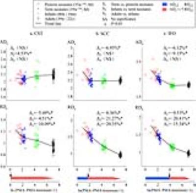
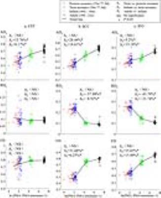
In CST, unchanged/increased ADe and decreased RDe are observed from preterm neonates to adults periods. In SCC and IFO, two change patterns of ADe and RDe are found (Figure 1). These results reveal the asynchrony of myelination across white matter structures. In the intra-axonal compartment, ADa and FD increase with age, while RDa decreases with age, especially from term neonates to adults periods (Figure 2). The trend line of ADa is not always same with FD. This indicates that ADa contains more information associated with axonal development, besides the axon organization.
DISCUSSION
Development processes in human brain white matter involve axon organization, premyelination (membrane proliferation), and myelination 1, 4, 8. The proliferation and growth of gilial cells and the increase of myelin sheath mainly lead to the structural changes in the extra-axonal space, which may be reflected by ADe and RDe. Premyelination leads to changes of diffusivities in almost all the directions, while myelionation mainly cause alterations in the radial direction 8. According to the combination of extra-axonal directional diffusivities, CST, SCC, and IFO start their myelination during the prenatal period, after birth, and at around postnatal one year, respectively. This is in agreement with the myelination sequence 8. Developmental changes of the fiber dispersion and intra-axonal diffusivities are observed during pre-myelination and myelination periods. These results reveal that the axonal development exists in pre-myelination and myelination periods. Increased ADa is related to the axonal organization 6. The results in this study demonstrated that changes of ADa can be also influenced by other factors associated with axon growth.CONCLUSION
Directional diffusivities in intra-axonal and extra-axonal compartments are valuable for evaluating the axonal development in vivo, as well as the pre-myelination and myelination processes.Acknowledgements
This work was supported by the grant from the National Natural Science Foundation of China (No.81171317, 81471631), the National Key Research and Development Program of China (2016YFC0100300), and the 2011 New Century Excellent Talent Support Plan from the Ministry of Education of China (NCET-11-0438).References
1. Dubois J, Dehaene-Lambertz G, Kulikova S, et al. The early development of brain white matter: a review of imaging studies in fetuses, newborns and infants. Neuroscience 2014;276:48-71.
2. Paus T. Growth of white matter in the adolescent brain: myelin or axon? Brain and Cognition 2010;72:26-35.
3. Paus T, Zijdenbos A, Worsley K, et al. Structural maturation of neural pathways in children and adolescents: in vivo study. Science 1999;283:1908-1911.
4. Qiu A, Mori S, Miller MI. Diffusion tensor imaging for understanding brain development in early life. Annual Review of Psychology 2015;66:853-876.
5. Fieremans E, Jensen JH, Helpern JA. White matter characterization with diffusional kurtosis imaging. Neuroimage 2011;58:177-188.
6. Jelescu IO, Veraart J, Adisetiyo V, et al. One diffusion acquisition and different white matter models: how does microstructure change in human early development based on WMTI and NODDI? Neuroimage 2015;107:242-256.
7. Oishi K, Mori S, Donohue PK, et al. Multi-contrast human neonatal brain atlas: application to normal neonate development analysis. Neuroimage 2011;56:8-20.
8. Dubois J, Dehaene-Lambertz G, Perrin M, et al. Asynchrony of the early maturation of white matter bundles in healthy infants: quantitative landmarks revealed noninvasively by diffusion tensor imaging. Human Brain Mapping 2008;29:14-27.
Figures


