4057
STRUCTURAL CONNECTIVITY ABNORMALITIES UNDERLYING COGNITIVE IMPAIRMENT IN PEDIATRIC MULTIPLE SCLEROSIS1Neuroimaging Research Unit, San Raffaele Scientific Institute, Vita-Salute San Raffaele University, Milan, Italy, 2Department of Neurology, San Raffaele Scientific Institute, Vita-Salute San Raffaele University, Milan, Italy, 3Multiple Sclerosis Center, Ospedale di Gallarate, Gallarate, Italy, 4Fondazione "Istituto Neurologico Casimiro Mondino", Pavia, Italy, 5Multiple Sclerosis Center, Spedali Civili di Brescia, Brescia, Italy, 6Department of Neurology, University of Florence, Florence, Italy, 7Department of Neuroradiology, San Raffaele Scientific Institute, Vita-Salute San Raffaele University, Milan, Italy
Synopsis
In this study, diffusion tensor (DT) magnetic resonance imaging (MRI) was applied to describe brain structural network architecture and connectivity abnormalities underlying cognitive dysfunction in 53 pediatric multiple sclerosis (MS) patients in comparison to 26 age- and gender-matched healthy controls (HC). Global and local network analyses were performed to assess between-group differences of connectivity metrics and cortical hubs. Cognitive impairment in pediatric MS patients seemed to be mainly associated to a reduced strength of connections of structural hubs and loss of efficiency in information transmission.
Purpose
A high proportion of patients with pediatric multiple sclerosis (MS) suffers from cognitive deficits with a prominent involvement of linguistic abilities in addition to memory, attention, and executive functions.1-3 However, the factors associated with cognitive impairment in these patients remain largely unexplored.4,5 Aim of this study was to describe brain structural network architecture in pediatric MS patients applying graph-analysis and to detect structural connectivity abnormalities underlying cognitive dysfunction across the different cognitive domains.Methods
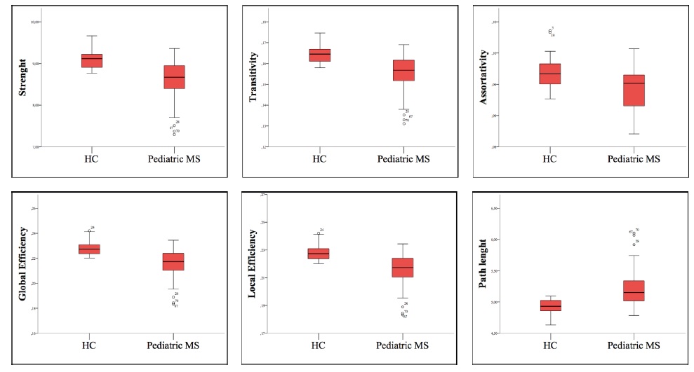
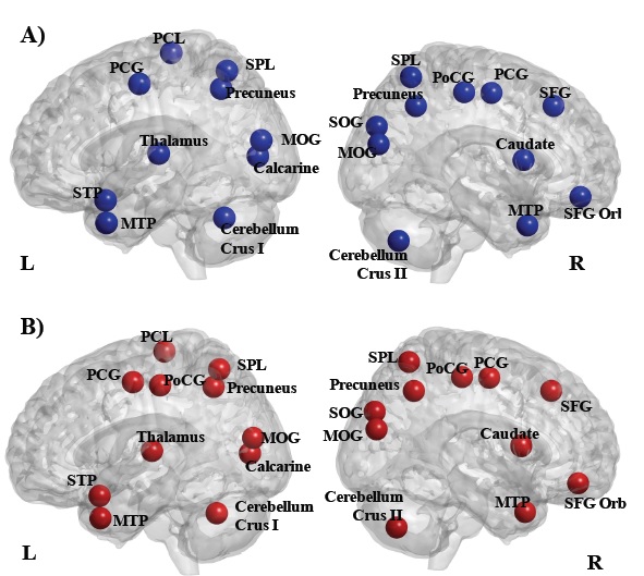
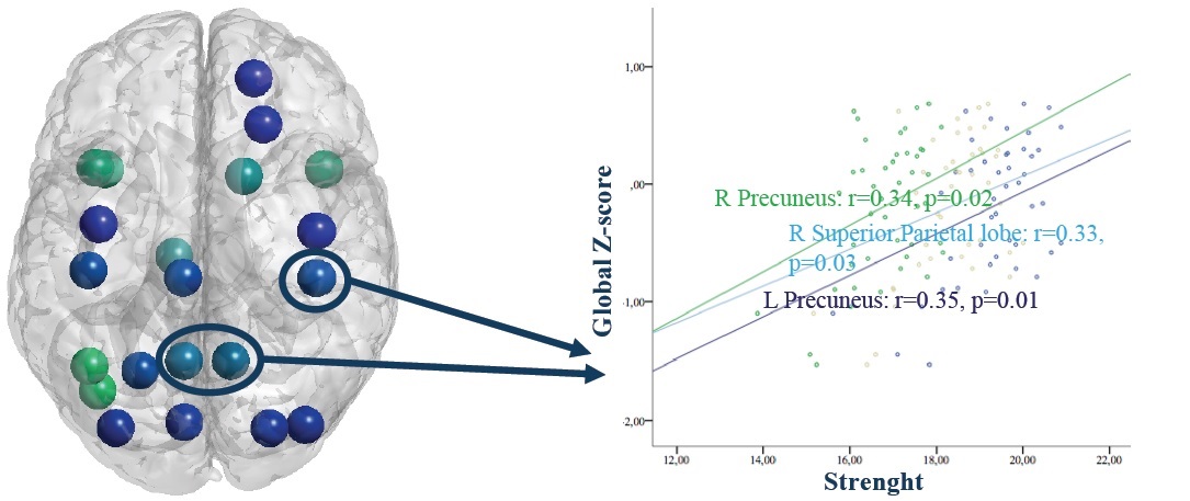
Diffusion tensor and dual echo turbo spin echo MRI scans were obtained from 53 pediatric MS patients and 26 age- and gender-matched healthy controls (HC), using a 3.0 T scanner. A first pre-processing involving distortions and motion correction (topup tool), tensor estimation and non-linear registration into the MNI space (fnirt tool) was performed. Between-group differences of global network were investigated evaluating the strength, assortativity, transitivity, global efficiency, local efficiency, average and path length (Figure 1). Local network connectivity metrics were investigated through the assessment of nodal strength, betweeness centrality and cortical hubs (Figure 2).6 Partial correlations between network metrics and Z-scores for each of cognitive domains and a global Z-score of cognitive function controlling for age and sex were evaluated.Results
All global network metrics differed significantly between pediatric MS patients and HC. Compared to HC, pediatric MS patients showed only an additional hub in the left post-central gyrus. A significant reduction of the strength in all network nodes identified as hubs was detected. Global cognitive function was positively correlated with the strength of connections of hubs located in the right superior parietal lobe and bilateral precuneus (Figure 3). Impairment in language functions and verbal memory were significantly related to a reduction in strength of the hubs located in frontal and temporal lobes, while visual-spatial memory, attention and information processing speed impairment were associated to a reduced strength in several hubs located in frontal, parietal and occipital lobes.Conclusions
Abnormalities of global network metrics occur in pediatric MS patients with limited differences in hubs distribution, indicating a partial preservation of brain network architecture. Cognitive impairment is mainly associated to a globally reduced strength of connections of the nodes identified as hubs, likely due to diffuse normal-appearing white matter damage, more than to a local damage, resulting in alteration and loss of efficiency in information transmission.Acknowledgements
No acknowledgement found.References
1. Weisbrot D, Charvet L, Serafin D, et al. Psychiatric diagnoses and cognitive impairment in pediatric multiple sclerosis. Mult Scler. 2014; 20(5):588-93.
2. Amato MP, Krupp LB, Charvet LE, et al. Pediatric multiple sclerosis: Cognition and mood. Neurology. 2016; 87:S82-7.
3. Lori S, Portaccio E, Zipoli V, et al. Cognitive impairment and event-related potentials in paediatric multiple sclerosis: 2-year study. Neurol Sci. 2011; 32(6):1043-6.
4. Charvet L, Cersosimo B, Schwarz C, et al. Behavioral Symptoms in Pediatric Multiple Sclerosis: Relation to Fatigue and Cognitive Impairment. J Child Neurol. 2016; 31(8):1062-7.
5. Banwell B, Arnold DL, Tillema JM, et al. MRI in the evaluation of pediatric multiple sclerosis. Neurology. 2016; 87:S88-96.
6. Filippi M, van den Heuvel MP, Fornito A, et al. Assessment of system dysfunction in the brain through MRI-based connectomics. Lancet Neurol. 2013; 12:1189-99.
Figures