4054
Brain sodium concentrations in healthy subjects are constant over time: a 3-year longitudinal 23Na MRI study at 3T1Aix-Marseille Univ, CNRS, CRMBM, Marseille, France, 2Aix-Marseille Univ, APHM, Hopital de la Timone, CEMEREM, Marseille, France, 3Aix Marseille Univ, APHM, Hôpital de la Timone, Pôle de Neurosciences Cliniques, Service de Neurologie, Marseille, France
Synopsis
Longitudinal evaluation of brain sodium concentration in physiological conditions
Introduction
Sodium (23Na) MRI provides a unique, non-invasive way to detect and quantify in vivo sodium concentrations based on the magnetic properties of the 23Na nucleus. In the human brain, sodium is distributed in two compartments (approximately 15mM in the intracellular and 140mM in the extracellular compartment) and any impairment of energy metabolism or achievement of the cell membrane integrity leads to an increase of the intracellular sodium concentration and consequently of the Total Sodium Concentration (TSC), which is the averaged sodium concentration between the intra- and extracellular compartments. The feasibility of sodium MRI to examine the human brain was first demonstrated in the 1980’s (1) and the recent advances in scanner technology (ultra high field, fast and strong gradients) and sequences design (ultra short echo time sequences) allowed the application of sodium MRI in clinical research. A number of studies have recently demonstrated the potential of brain sodium MRI to non-invasively detect total sodium accumulations in neurological disorders and diseases such as stroke (2-4), brain tumors (5-6), Huntington’s disease (7), Alzheimer disease (8) and multiple sclerosis (9-13). All these studies showed a sodium accumulation compared to healthy controls. Nevertheless, any study has explored the physiological variation of sodium concentration over time in healthy people. This is an important prerequisite for future longitudinal studies in neurological disorders. The aim of the present study was to assess the longitudinal variations of brain sodium concentrations in healthy subjects during a three-year follow up.Methods
MR scans were performed on the same 3T Verio system holding multi-nuclear options (Siemens, Erlangen, Germany) in 22 healthy controls. 23Na MRI was acquired using a double-tuned 23Na-1H volume head coil (Rapid Biomedical, Rimpar, Germany) and a 3D density-adapted radial projection reconstruction pulse sequence (TE=200μs, TR=120ms, 17000 projections and 369 samples per projection, 3.6mm3 isotropic resolution, acquisition time = 34min) with two tubes filled with 50 mM of sodium placed in the FOV to serve for external references. High-resolution proton MRI 3D-MPRAGE (TR=2300ms, TE=3ms, TI=900ms, 160 slices, 1mm3 isotropic resolution) was obtained using a 32-element 1H head coil (Siemens). The optimized post-processing pipeline is described in Figure 1 and allowed to obtain total sodium concentration (TSC) from grey matter and white matter. Statistical analysis was performed using non-parametric tests.Results
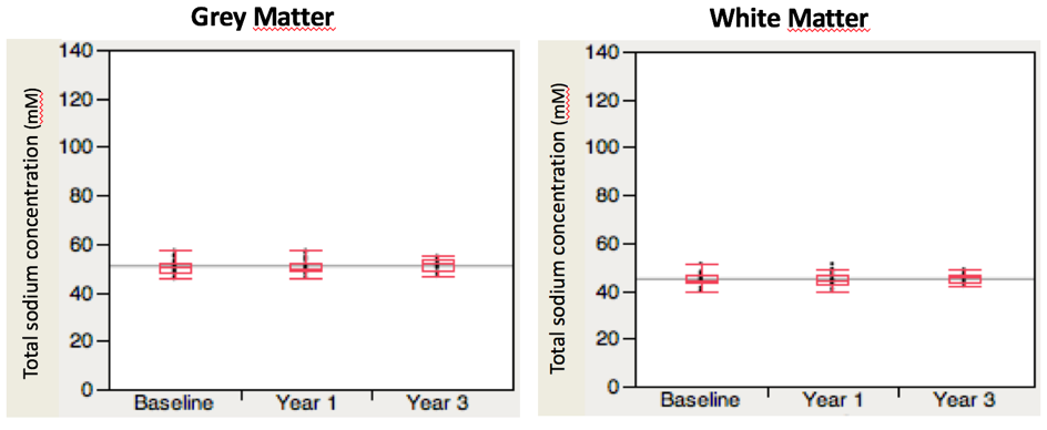
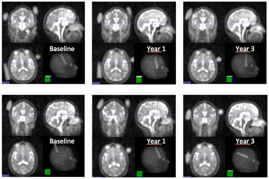
Twenty-two healthy volunteers were enrolled for this longitudinal study (9 women and 13 men). The mean age was 33 (±11) yo. At baseline, the mean grey matter total sodium concentration (GM-TSC) was 50.62 (±2.92) mM wet tissue volume and the mean white matter total sodium concentration (WM-TSC) was 44.66 (±2.60) mM wet tissue volume. Twenty volunteers were scanned at one year. The mean GM-TSC was 50.56 (±3.04) mM wet tissue volume and the mean WM-TSC was 44.64 (±2.69) mM wet tissue volume. Eleven volunteers were scanned at three years. The mean GM-TSC was 51.39 (±2.68) mM wet tissue volume and the mean WM-TSC was 45.58 (±2.08) mM wet tissue volume. These concentrations were not different over time (p=0.76 for GM-TSC and p=0.56 for WM-TSC) (Figure 2). An example of sodium images over time for two controls is shown in Figure 3.Discussion
This longitudinal sodium MRI study has demonstrated stability and reproducibility of sodium concentration in healthy subjects over three years. This finding is of importance as reproducibility over several years of sodium MRI in physiological conditions strengthens results from previous studies showing sodium accumulation in neurological diseases compared to controls (14). Furthermore, while these previous studies mainly used cross-sectional design, the present report is an important prerequisite for future longitudinal studies focusing in neurological disorders where a variation of sodium concentration over time is expectedAcknowledgements
No acknowledgement found.References
(1) Hilal SK, Maudsley AA, Ra JB, Simon HE, Roschmann P, Wittekoek S, et al. In vivo NMR imaging of sodium-23 in the human head. J. Comput. Assist. Tomogr. 1985; 9: 1–7.
(2) Boada FE, Qian Y, Nemoto E, Jovin T, Jungreis C, Jones SC, et al. Sodium MRI and the assessment of irreversible tissue damage during hyper-acute stroke. Transl. Stroke Res. 2012; 3: 236–245.
(3) Hussain MS, Stobbe RW, Bhagat YA, Emery D, Butcher KS, Manawadu D, et al. Sodium imaging intensity increases with time after human ischemic stroke. Ann. Neurol. 2009; 66: 55–62
(4) Thulborn KR, Davis D, Snyder J, Yonas H, Kassam A. Sodium MR imaging of acute and subacute stroke for assessment of tissue viability. Neuroimaging Clin. N. Am. 2005; 15: 639–653, xi–xii.
(5) Ouwerkerk R, Bleich KB, Gillen JS, Pomper MG, Bottomley PA. Tissue sodium concentration in human brain tumors as measured with 23Na MR imaging. Radiology 2003; 227: 529–537.
(6) Thulborn KR, Lu A, Atkinson IC, Damen F, Villano J. Quantitative Sodium MR Imaging and Sodium Bioscales for the Management of Brain Tumors. Neuroimaging Clin. N. Am. 2009; 19: 615–624.
(7) Reetz K, Romanzetti S, Dogan I, Saß C, Werner CJ, Schiefer J, et al. Increased brain tissue sodium concentration in Huntington’s Disease - a sodium imaging study at 4 T. NeuroImage 2012; 63: 517–524.
(8) Mellon EA, Pilkinton DT, Clark CM, Elliott MA, Witschey WR, Borthakur A, et al. Sodium MR imaging detection of mild Alzheimer disease: preliminary study. AJNR Am. J. Neuroradiol. 2009; 30: 978–984
(9) Inglese M, Madelin G, Oesingmann N, et al. Brain tissue sodium concentration in multiple sclerosis: a sodium imaging study at 3 tesla. Brain. 2010;133:847–857.
(10) Zaaraoui W, Konstandin S, Audoin B, et al. Distribution of brain sodium accumulation correlates with disability in multiple sclerosis: a cross-sectional 23Na MR imaging study. Radiology. 2012;264:859–867.
(11) Paling D, Solanky BS, Riemer F, et al. Sodium accumulation is associated with disability and a progressive course in multiple sclerosis. Brain. 2013;136:2305–2317.
(12) Maarouf A, Audoin B, Konstandin S, et al. Topography of brain sodium accumulation in progressive multiple sclerosis. Magma N Y N. 2014;27:53–62.
(13) Petracca M, Vancea RO, Fleysher L, Jonkman LE, Oesingmann N, Inglese M. Brain intra- and extracellular sodium concentration in multiple sclerosis: a 7 T MRI study. Brain. 2016 Mar;139(Pt 3):795-806.
(14) Shah NJ, Worthoff WA, Langen K-J. Imaging of sodium in the brain: a brief review. NMR Biomed. 2016; 29(2):162–174.
Figures