4051
LONGITUDINAL ASSESSMENT OF LARGE-SCALE BRAIN FUNCTIONAL NETWORKS IN PATIENTS WITH MS: RELATIONSHIP WITH CLINICAL DISABILITY AND COGNITIVE IMPAIRMENTPaola Valsasina1, Maria Assunta Rocca1, Fiammetta Pirro1, Annalisa Colombi1, Elisabetta Pagani1, Ermelinda De Meo1, Bruno Colombo2, Paolo Preziosa1, Vittorio Martinelli2, Giancarlo Comi2, Andrea Falini3, and Massimo Filippi1
1Neuroimaging Research Unit, San Raffaele Scientific Institute, Vita-Salute San Raffaele University, Milan, Italy, 2Department of Neurology, San Raffaele Scientific Institute, Vita-Salute San Raffaele University, Milan, Italy, 3Department of Neuroradiology, San Raffaele Scientific Institute, Vita-Salute San Raffaele University, Milan, Italy
Synopsis
Aim of this study was to investigate the temporal evolution of resting state (RS) functional connectivity (FC) in patients with multiple sclerosis (MS) and its correlation with clinical and cognitive worsening. The predictive value of baseline functional network measures on the worsening of clinical disability/cognitive impairment was also explored. No significant RS FC changes were detected in healthy controls, while MS patients showed a complex pattern of longitudinal changes in the different networks, with a trend towards an increase (or stability) of RS FC in clinically stable MS patients, and a decrease of RS FC in clinically worsened MS patients.
Purpose
Resting state (RS) functional MRI (fMRI) allows to investigate intrinsic, synchronized brain activity across the whole brain and to measure the degree of functional correlations between different cerebral regions [1]. RS functional connectivity (FC) studies in multiple sclerosis (MS) detected a consistent reduction of cortical/subcortical RS FC compared with control subjects, which was clinically relevant. However, little is known about the temporal evolution of network RS FC and the association between network abnormalities and the development of physical and cognitive disability. Aim of this study was to investigate the temporal evolution of RS FC in patients with MS and its correlation with clinical and cognitive worsening.Methods
Using a 3T Philips scanner, dual echo, 3D T1-weighted, diffusion tensor and RS fMRI scans were obtained at baseline and follow-up (median time=3.6 years) from 56 right-handed MS patients and 24 matched healthy controls (HC). Patients also underwent clinical and neuropsychological evaluations at both time points. Based on Expanded Disability Status Scale (EDSS) score [2] modifications at follow-up, MS patients were classified as clinically stable or worsened. Seed-voxel RS FC was performed using seven major cortical/subcortical seeds (posterior cingulum, inferior parietal cortex, cuneus, postcentral gyrus, cerebellum, thalamus, and amygdala) described by Tomasi et al. [3]. Fractional anisotropy between regions of each functional network was also measured. RS FC longitudinal changes were compared between HC, clinically stable and worsened MS patients. Multivariable logistic models investigated the predictive role of clinical and RS FC variables on neurological/cognitive deterioration.Results
At follow-up, 11 MS patients (20%) were clinically worsened and 6 developed cognitive impairment. Clinically worsened MS patients had a higher EDSS and lower normalized brain volume at baseline than clinically stable MS patients. Clinically worsened MS patients had also a higher rate of new T2 and T1 lesion formation and a higher rate of brain atrophy development than clinically stable MS patients. Global functional and structural connectivity was lower in HC vs MS, and in worsened vs stable MS patients at both time points. RS FC remained stable over time in HC, while it increased (or remained stable) in clinically stable MS patients and decreased in clinically worsened patients, as shown in Figures 1 and 2. Structural connectivity decreased over time in both MS subgroups. A lower baseline thalamic and default-mode network RS FC predicted neurological deterioration at follow-up (p=range 0.004-0.04). Decreased RS FC in these same networks was associated with a worse cognitive performance at follow-up.Conclusions
Longitudinal modifications of RS FC occur in MS patients and differ from those of HC. Increased RS FC plays an adaptive role, probably in response to accumulation of structural damage. Such an increased RS FC in a given brain network in MS patients might protect from the onset of clinical deficits and cognitive impairment. Conversely, a distributed reduction of RS FC in clinically worsened and/or cognitively deteriorating MS patients is likely to reflect the exhaustion of brain functional reserve.Acknowledgements
This study was partially supported by a grant from FISM 2014/R/7.References
[1] Biswal B.B., et al. PNAS 2010; 107:4734-9. [2] Kurtzke J.F. Neurology 1983; 33:1444-52. [3] Tomasi D., et al. Cereb Cortex 2011; 21:2003-13.Figures
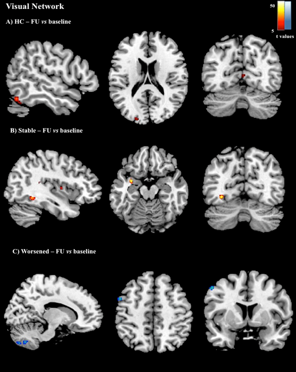
Within-group comparisons
for the visual network RS FC between baseline and FU assessment (foci of
increased RS FC at FU are indicated in red, foci of decreased FS FC are coded
in blue) (p<0.001, uncorrected).
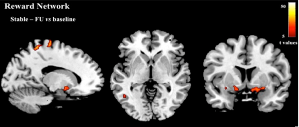
Within-group comparisons
for the reward network RS FC between baseline and FU assessment (foci of
increased RS FC at FU are indicated in red) (p<0.001, uncorrected).