4049
Myelin water imaging provides evidence of long-term remyelination and neuroprotection in Alemtuzumab treated multiple sclerosis patients1Radiology, University of British Columbia, Vancouver, BC, Canada, 2Pathology & Laboratory Medicine, University of British Columbia, Vancouver, BC, Canada, 3Medicine (Neurology), University of British Columbia, Vancouver, BC, Canada, 4Physics and Astronomy, University of British Columbia, Vancouver, BC, Canada
Synopsis
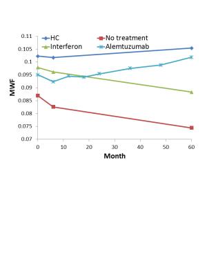
To test the potential neuroprotective and reparative properties of alemtuzumab (a highly effective disease modifying therapy for relapsing remitting MS), we used myelin water imaging to measure myelination in MS patients treated with either alemtuzumab, interferon, or no treatment. NAWM MWF showed a steady 4% increase in alemtuzumab-treated subjects whereas MWF in subjects treated with interferon or without treatment decreased by 10% over 5 years. Myelin recovery following treatment with alemtuzumab supports previous clinical trial findings, provides understanding of the biological mechanisms underlying observed clinical improvement and demonstrates that MWF is a powerful biomarker for neuroprotection and repair in MS.
Purpose
Alemtuzumab is a highly effective disease modifying therapy for relapsing and remitting multiple sclerosis (RRMS)1. To test the potential neuroprotective and reparative properties of the drug, we used myelin water imaging2 to measure myelination in vivo in MS patients treated with either alemtuzumab, interferon, or no treatment. Changes in myelin water fraction (MWF, a marker of myelin3) within normal-appearing white matter (NAWM) were monitored over 5 years.Methods
Thirty-one RRMS patients were imaged on a 3T Philips MR scanner. Twenty patients (3M/17F, mean age=35, median EDSS=3.5) were treated with alemtuzumab at baseline and 12 months, and scanned every 6 months for 2 years and then yearly until 4 or 5 years. Five patients (3M/2F, mean age=32, median EDSS=1) were treated with interferon subcutaneously 3 times per week and scanned at baseline, 6 months and ~5 years. Six patients (1M/5F, mean age=41, median EDSS=2) did not undergo treatment and were scanned at baseline, 6 months and ~5 years. Four healthy controls (1M/3F, mean age=43) were also scanned at baseline, 6 months and ~5 years. Scans included a 32 spin-echo T2 relaxation sequence (TE=10ms, TR=1000ms, voxel=1x1x5mm3, 7 slices) and a 3D volumetric T1 turbo field echo sequence (TE=3.6ms, TR=8ms, voxel=1x1x1mm3, TFE factor=200, flip angle=15o). T2 distributions were calculated for every voxel using a modified Extended Phase Graph algorithm combined with regularized non-negative least squares, flip angle optimization and spatial regularization4-6. NAWM was segmented from the T1 volumetric scan using FAST7. The mean MWF was measured in NAWM at all time-points up to year 5.Results
MWF results from NAWM are shown in Figure 1. MWF in NAWM showed a steady 4% increase in subjects treated with alemtuzumab (baseline=0.095; year 4=0.099; p=0.03) whereas the MWF in subjects treated with interferon (baseline=0.098; year 5=0.088; p=0.15) or without treatment (baseline=0.087; year 5=0.078; p=0.03) decreased by 10% over 5 years. MWF in healthy controls remained stable over 5 years (baseline=0.102, year 5=0.105; p=0.49) (Note that only 2 subjects treated with alemtuzumab had year 5 scans so the comparison was made between baseline and year 4.)Discussion
NAWM is known to be damaged in MS with histological evidence of demyelination8. MS treatment with interferons has shown some improvement in MRI outcomes such as a lower rate of brain atrophy9, increased magnetization transfer ratio (MTR) in lesions10 and increased NAA concentration as measured by 1H-MRS11. However, none of these measures are specific to myelin or show reversal of brain damage due to MS. Alemtuzumab is a newer disease modifying therapy shown to be clinically more effective at treating relapses than interferon over 2 years1. A 3 year advanced MRI study showed a stabilization of the NAWM MTR in subjects treated with alemtuzumab (-0.02 pu/year, p = 0.51) compared to a decline observed in untreated subjects (-0.12 pu/year, p = 0.004)12. The present study shows a steady improvement of the MWF in subjects with MS who are treated with alemtuzumab.Conclusions
Alemtuzumab has shown durable efficacy on clinical outcomes (such as disability improvement, sustained reduction in relapses and brain volume loss) during follow-up of the CARE-MS clinical trials1. Myelin recovery following treatment with alemtuzumab supports previous clinical trial findings and provides further understanding of the biological mechanisms underlying observed clinical improvement. The sensitive, specific and quantitative nature of myelin water imaging allowed detection of a treatment effect in a very small group, outside of areas of acute damage. This study demonstrates that MWF is a powerful biomarker for neuroprotection and repair in MS. This is the first long-term observational study showing evidence supportive of remyelination with a high efficacy therapy.Acknowledgements
This study was supported by Sanofi-Genzyme Corporation. We would like to thank the patients, volunteers and technologists. We acknowledge the continuing support from Philips Healthcare.References
1. Coles AJ, Twyman CL, Arnold DL, Alemtuzumab for patients with relapsing multiple sclerosis after disease-modifying therapy: a randomised controlled phase 3 trial. Lancet 2012; 380, 1829-1839.
2. MacKay A, Whittall K, Adler J, et al. In vivo visualization of myelin water in brain by magnetic resonance. Magn Reson Med 1994; 31(6): 673-677.
3. Laule C, Kozlowski P, Leung E, et al. Myelin water imaging of multiple sclerosis at 7 T: correlations with histopathology. Neuroimage 2008; 40(4): 1575-1580.
4. Prasloski T, Mädler B, Xiang Q-S, et al. Applications of stimulated echo correction to multicomponent T2 analysis. Magn Reson Med 2012; 67: 1803–1814.
5. Whittall KP and MacKay AL. Quantitative interpretation of NMR relaxation data. J Magn Reson 1989; 84: 134–152.
6. Yoo Y and Tam R. Non-local spatial Regularization of MRI T2 relaxation images for myelin water quantification. In: MICCAI, 2013, 8149, pp.614-621.
7. Zhang, Y. and Brady, M. and Smith, S. Segmentation of brain MR images through a hidden Markov random field model and the expectation-maximization algorithm. IEEE Trans Med Imag, 2001; 20(1):45-57.
8. Allen IV and McKeown SR. A histological, histochemical and biochemical study of the macroscopically normal white matter in multiple sclerosis. J Neurol Sci 1979; 41: 81-91.
9. Zivadinov R, Locatelli L, Cookfair D, et al. Interferon beta-1a slows progression of brain atrophy in relapsing-remitting multiple sclerosis predominantly by reducing gray matter atrophy. Mult Scler 2007; 13(4): 490-501.
10. Kita M, Goodkin DE, Bacchetti P, et al. Magnetization transfer ratio in new MS lesions before and during therapy with IFNβ-1a. Neurology 2000; 54(9): 1741-1745.
11. Sarchiellia P, Presciuttib O, Tarduccib R, et al. 1H-MRS in patients with multiple sclerosis undergoing treatment with interferon β-1a: results of a preliminary study. J Neurol Neurosurg Psychiatry 1998; 64: 204-212.
12. Button T, Altmann D, Tozer D, et al. Magnetization transfer imaging in multiple sclerosis treated with alemtuzumab. Mult Scler J 2013; 19(2): 241-244.
