4037
Ultra-high-bandwidth, high-resolution MRI of fast relaxing spins1University and ETH Zurich, Zürich, Switzerland
Synopsis
High resolution MRI of fast relaxing spins in human sets strong requirements on hardware and pulse sequences. Indeed, the acquisition duration is limited by the relaxation time of the protons of interest and high resolution can only be achieved with large magnetic field gradients. Also when large field-of-view is required, additional challenges appear. The sequence and receive hardware needs to be adapted to larger bandwidth. In this work, we image short T2 compounds in human scanners with gradient strengths up to 200 mT/m and bandwidth up to 1.63 MHz.
Purpose
High resolution MRI of fast relaxing spins in human sets strong requirements on hardware and pulse sequences. Indeed, the encoding duration is limited by the relaxation time of the protons of interest, and high resolution can only be achieved with large magnetic field gradients. Apart from the challenges for the gradient hardware, the sequence and receive hardware needs to be capable to handle the larger bandwidth. In this work, we image short T2 compounds in human scanners with gradient strengths up to 200 mT/m and bandwidth up to 1.63 MHz using a PETRA sequence1 combined with modulated excitation pulses2.Method
Hardware:
- Scanner: Philips 3T achieva with custom-made insert gradient coil with maximum strength 200 mT/m.
- RF: symmetrically biased TR-switches4, 1H-free coils.
- Pulse sequence: Modulated pulses (hyperbolic-secant of 10 us) were used in combination with PETRA in order to achieve a homogeneous sample excitation and recover the data missed during the T/R-switch dead time.
- Acquisition : standalone National Instrument spectrometer5 allowing acquisition with bandwidths up to 4 MHz.
- Reconstruction: Algorithm proposed by Li et al.6 was implemented in Matlab.
- Resolution phantoms:
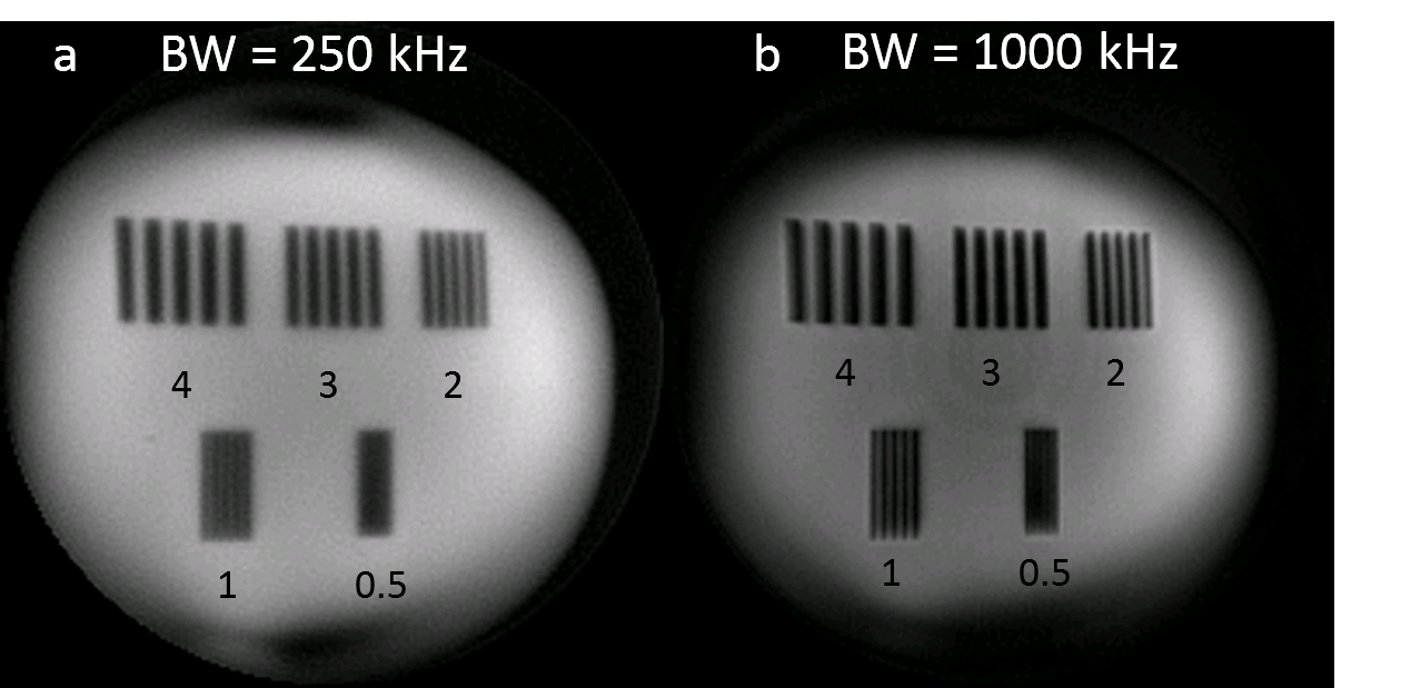
1) Rubber: T2 ~= 180 us (exponential fit of FID), resolution stripes: 4, 3, 2, 1 and 0.5 mm.
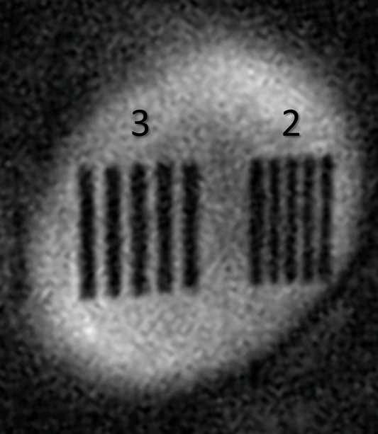
2) PMMA: T2 = a few tens of microseconds7, resolution stripes: 3 and 2 mm.
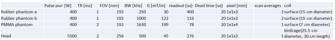
All other imaging parameters are given in table 1.
Results
Figure 1: Demonstrates the feasibility of imaging short-T2 protons at high resolution and high bandwidth. The resolution is strongly improved at 1 MHz compared to 250 kHz bandwidth and approaches the nominal pixel size of 1 mm. Remaining deformation is due to uncorrected gradient non-linearity.
Figure 2: Demonstrates that a resolution of ~2 mm can be obtained on hard, usually invisible, polymers such as PMMA with relaxation times of a few tenths of microseconds.
Figure 3: High gradient, high bandwidth imaging is also feasible in-vivo. In this figure, fines structures down to 1 mm are well depicted in the nasal cavity. The ring-like artifact apparent in the brain is associated to spurious signal from transmit-receive switching.
Discussion
We demonstrated the feasibility of high resolution short-T2 imaging at ultra-high bandwidth up to 1.63 MHz both in phantoms and in-vivo, employing a dedicated insert gradient, optimized sequences, and RF hardware.
Further improvements in image quality are expected by removing spurious signal on the receive chain and by the use of coil-arrays to increase the signal-to-noise ratio.
Finally, this work demonstrates that the study of fast-relaxing spins in the human body will greatly profit from state-of-the-art hardware and pulse sequences that allow the acquisition of high bandwidth images. To this end, the creation of contrast by isolation of the signal of interest will be of high importance.
Acknowledgements
No acknowledgement found.References
1. Grodzki DM, Jakob PM, Heismann B. Ultrashort echo time imaging using pointwise encoding time reduction with radial acquisition (PETRA). Magn Reson Med. 2012;67:510-518. doi:10.1002/mrm.23017.
2. Schieban K, Weiger M, Hennel F, Boss A, Pruessmann KP. ZTE Imaging With Enhanced Flip Angle Using Modulated Excitation. 2014;00:1-10. doi:10.1002/mrm.25464.
3. Brunner DO, Furrer L, Weiger M, et al. Symmetrically Biased T/R Switches for NMR and MRI with Microsecond Dead Time. J Magn Reson. 2016;263:147-155. doi:10.1016/j.jmr.2015.12.016.
4. Weiger M, Brunner DO, Dietrich BE, Müller CF, Pruessmann KP. ZTE imaging in humans. Magn Reson Med. 2013;70(2):328-332. doi:10.1002/mrm.24816.
5. Li C, Magland JF, Zhao X, Seifert AC, Wehrli FW. Selective In Vivo Bone Imaging with Long- T 2 Suppressed PETRA MRI. 2016;00:1-9. doi:10.1002/mrm.26178.
6. Davies GR, Lurie DJ, Hutchison JMS, McCallum SJ, Nicholson I. Continuous-Wave Magnetic Resonance Imaging of Short T2 Materials. J Magn Reson. 2001;148(2):289-297. doi:10.1006/jmre.2000.2245.
Figures