4035
Fast, Volumetric and Silent Multi-contrast Zero Echo Time Imaging1Technical University Munich, Garching, Germany, 2GE Global Research, Garching, Germany
Synopsis
The current work aims to provide a volumetric, fast and silent method for quantitative T1 mapping with Zero Echo Time (ZTE) imaging, and generate multiple T1-weighted images at virtual inversion times. By designing an interleaved radial trajectory for ZTE, and constraining the temporal behavior of the signal with low-dimensional subspace and spatiotemporal low rank regularization, we conducted a volumetric T1 mapping in 2 minutes with acoustic noise only 1.1dB higher than scanner background.
Purpose
Zero
echo time (ZTE) imaging has many favorable features, as it is distortion free,
silent and can provide isotropic resolutions, however with poor image contrast1. The aim of this work is to generate multiple T1 contrast images at virtual
inversion times (TIs) and quantitative T1 maps with magnetization prepared ZTE,
in clinical feasible time and low acoustic noise to enhance patient comfort. We
designed an interleaved radial trajectory to best elevate scan efficiency, and
implemented a low dimensional temporal subspace and low rank (LR) regularization
method2,3 for reconstruction. Methods
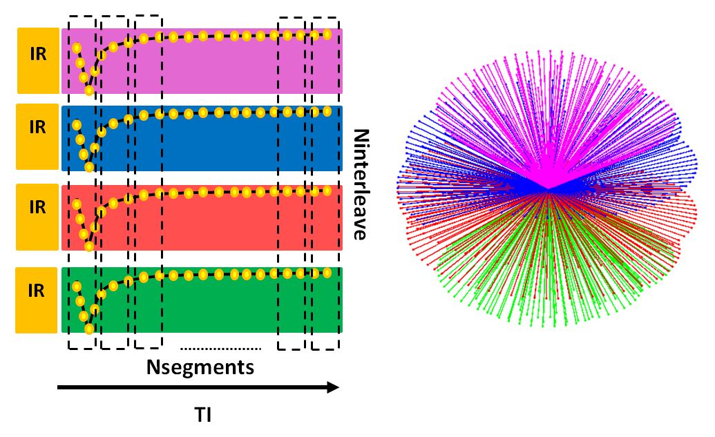
Sequence design: The radial trajectory is designed according to an interleaved Archimedean spiral trajectory4. The acquired data is segmented along the readout, and data segments with the same position in the readout (similar TI) from different interleaves are grouped together to generate undersampled images. The effective TI is defined as the center of the acquisition window for each segment. The trajectory of each interleave is shifted to guarantee that the undersampled volumes can uniformly cover the K space. The trajectory design is shown in Fig. 1.
Data acquisition: The experiment was conducted on a GE 3T MR750w scanner with a GEM head array coil (GE Healthcare, Waukesha, WI). A phantom (DiagnosticSonar, Livingston, UK) consisting of tubes with different T1 values were used in the experiment. An adiabatic inversion recovery (IR) prepared ZTE was conducted with FOV=21.6cm, flip angle=2°, readout BW = ±15.6 kHz, isotropic resolution of 3mm, and a waiting time of 1000ms to allow signal recovery between consecutive interleaves. The data acquisition began 40ms after the IR pulse and lasted 3000ms. Acoustic noise measurements were performed using a Bruel&Kjaer sound level meter equipped with MR compatible microphone, which was placed in-bore at scanner isocenter inside the head coil. An initial volunteer scan was also conducted with the same imaging protocol.
Data reconstruction: The compressed sensing reconstruction with low dimensional temporal subspace constraint and LR regularization was implemented3. The observed signal y(t) can be modeled as y=EXt, in which E is the encoding operator and Xt represents the temporal dynamic image series. The signal evolution is a function of tissue parameter (T1, proton density) and IR pulse flip angle (due to imperfect IR pulse), and can be approximated by temporal basis determined by the signal model. The LR regularization exploits spatiotemporal correlations and reduces the degree of freedoms in the subspace. In the current work we chose K=3 temporal coefficients, and segmented the data into 64 time points with different TIs.
Results
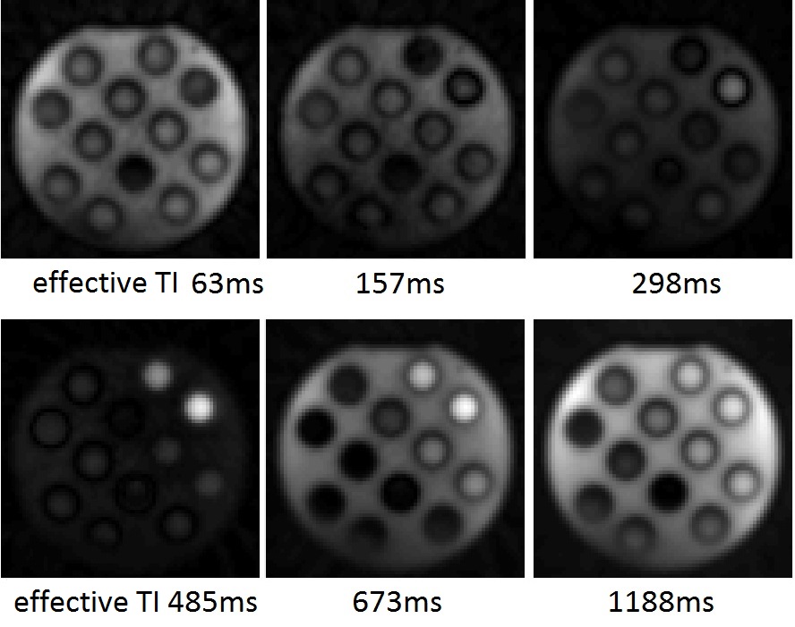
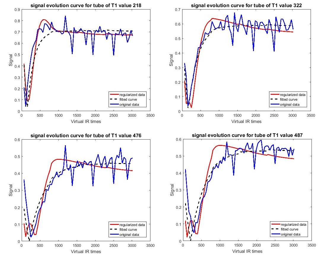
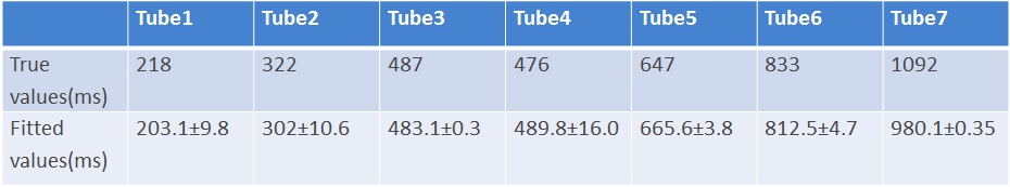
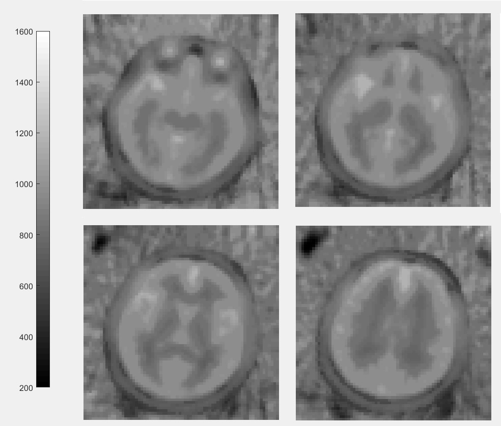
As shown in Figure 2, the temporal behavior of different T1 samples can be observed in reconstructed image series at different virtual TIs. Fig 3 shows the signal evolution before and after applying temporal subspace and LR regularization, and the fitted signal curve. Mean and standard deviation of the fitting results for each tube are shown in Table1. Compared to vendor provided ground truth T1 values, the fitting results are similar but with an underestimation of long T1 (tube7). T1 values consistent with literature were generated in gray and white matter area from the volunteer scan, and are shown in Figure 4. However, the acquisition and reconstruction parameters need further improvement to generate decent T1 mapping in vivo. The current experiment took less than 2 minutes, and the acoustic noise was 71.1dB, only 1.1dB higher than the background noise (70.0dB).Discussion
In this study we conducted volumetric, fast and silent T1 mapping and reconstructed multiple T1 contrast images at virtual TIs with IR prepared ZTE. The current method was validated in T1 phantom and initially validated in volunteer scanning, yet further improvement is necessary. Unlike Cartesian or spiral trajectory, radial trajectory has no defined contrast point as it constantly updating the center of K space. Previous studies5 used view-sharing for IR prepared radial sequence to solve the contradiction between image quality and contrast. In this work, we utilized an alternative method by having undersampled images acquired at similar TI times, and reduce undersampling artifacts by low-dimensional subspace and LR regularization.
Additionally, there are several limitations that need to be improved in next steps. First, there was an underestimation bias of long T1 values, which could be improved by a longer acquisition window to better capture the dynamic relaxation curve of long T1 samples. Second, the signal model does not consider inaccurate flip angle during ZTE readout which could also affect the signal evolution. Including the possible flip angle variations in the signal model could increase the accuracy in calculating the temporal basis.
Acknowledgements
This work was funded by the European Commission under Grant Agreement Number 605162.
References
1. Peter Börnert et al, ISMRM23(2015), 0510: Magnetization Prepared ZTE to address Multiple Diagnostic Contrasts
2. Jonathan I. Tamir et al, ISMRM23(2015), 3399: T2 Shuffling: Multicontrast 3D Fast Spin Echo Imaging
3.Jonathan I. Tamir et al, T2 Shuffling: Sharp, Multicontrast, Volumetric Fast Spin-Echo Imaging, MRM 2016, doi 10.1002/mrm.26102
4. Sam T.S. Wong et al, A Strategy for Sampling on a Sphere Applied to 3D Selective RF Pulse Design, MRM 1994, 32: 778–784
5. Steven Kecskemeti et al, MPnRAGE: A technique to simultaneously acquire hundreds of differently contrasted MPRAGE images with applications to quantitative T1 mapping, MRM 2016,75:1040–1053
Figures