4028
2D-UTE based pantomography with half-pulse excitation1Internal Medicine II, University Hospital of Ulm, Ulm, Germany, 2Department of Medical Physics, Memorial Sloan Kettering Cancer Center, New York, NY, United States, 3Dental Imaging, Sirona Dental Systems GmbH, Bensheim, Germany
Synopsis
Standard imaging methods in dental radiology are almost exclusively based on X-rays. Not only does magnetic resonance imaging (MRI) avoid ionizing radiation, but has also been shown to be superior in identifying carious lesions. In this work we present a 2D-UTE sequence with an echo time of 35µs, utilizing 240µs half-pulses to ensure reduced signal attenuation from tissues with short T2 relaxation during excitation. The feasibility of the method was demonstrated with the successful acquisition of in vivo MR pantomograms.
Introduction
X-ray based pantomography is the current standard for deriving overview scans of the jaw and teeth in dental routine. Considering the potential risk from the exposure to ionizing radiation and the only limited visualization of soft-tissue structures, alternative approaches for the generation of pantomograms are of interest. The application of MRI was so far limited by its rather poor performance in diagnosis of mineralized structures. Even though ultra-short echo time techniques like UTE, ZTE, and SWIFT have proven applicable to bone and tooth imaging, the intrinsic volumetric imaging approach is limiting their application for providing pantomograms at sufficiently high spatial resolution in clinically acceptable scan times. Translation to two-dimensional UTE imaging is limited by the strong signal decay due to the required slice-selective excitation.
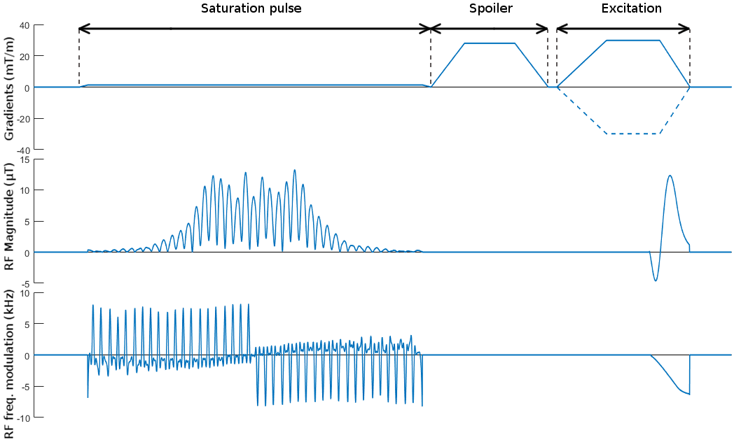
In this work 2D-UTE was combined with a short 240µs half-sinc pulse to ensure minimal signal decay during excitation, a quadratic phase saturation pulse for the suppression of remaining out-of-slice signal, and a ZTE-like reconstruction to cope with the transient phase of the FIR filter.
Methods
In half-sinc excitation, the excitation is split into subsequent acquisitions, each covering one half of excitation k-space, thus avoiding any rephasing gradient. The combination of both excitations, in theory, yields a perfect slice profile1.
To avoid severe signal decay during excitation, the length of the half-sinc pulse was chosen as short as possible for a given slice-thickness, which yielded playing out of the rf-waveform almost entirely on the ramp-down of the gradient. Distortions of the gradient waveform due to eddy-currents were considered during rf-waveform calculation and to ensure proper off-center slice profiles a modulation with $$$\exp\left({iz_{0} \gamma \int_{t}^{T}g(\tau)\text{d}\tau} \right)$$$was added to the pulse with $$$z_{0}$$$ and $$$g(\tau)$$$ being the off-center position and gradient waveform, respectively2.
Despite all these measures, the half-pulse approach remains quite susceptible to unaccounted gradient imperfections and off-resonances, yielding tissue excitation outside the intended slice. To counter these effects, a 8ms quadratic phase saturation pulse with TBP = 34, a slice thickness of 10mm and saturation bands of 5cm on either side of the slice was used3, followed by a spoiler gradient. A sequence diagram is provided in Figure 1.
All in vivo data was acquired with a 1.5T whole-body clinical imaging system (Achiva 1.5T, Philips Healthcare, Best, The Netherlands) using a four-element dental surface coil (NORAS, Hoechberg, Germany) with tune delay set to 10µs. The phantom images were acquired with a 3T whole-body clinical imaging system (Achiva 3T, Philips Healthcare, Best, The Netherlands). Acquisition parameters for in vivo images were as: slice thickness of 10mm, flip angle 15°, TR = 120ms, and 0.5x0.5mm² in-plane resolution with an echo time of TE=750µs (reference standard 2D-UTE) and TE=35µs (240µs half-pulse). The first 8 data points were corrupted by the transient phase of the FIR filter and a still partly detuned coil. Considering the strong over-sampling in the center of k-space and the very low gradient in the beginning of the trajectory, corrupted data points were removed and images reconstructed with compressed sensing technique4 applying a TV-filter for regularization.
Results
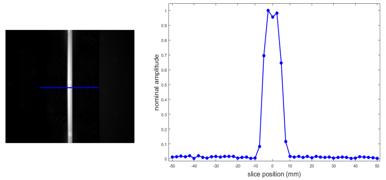
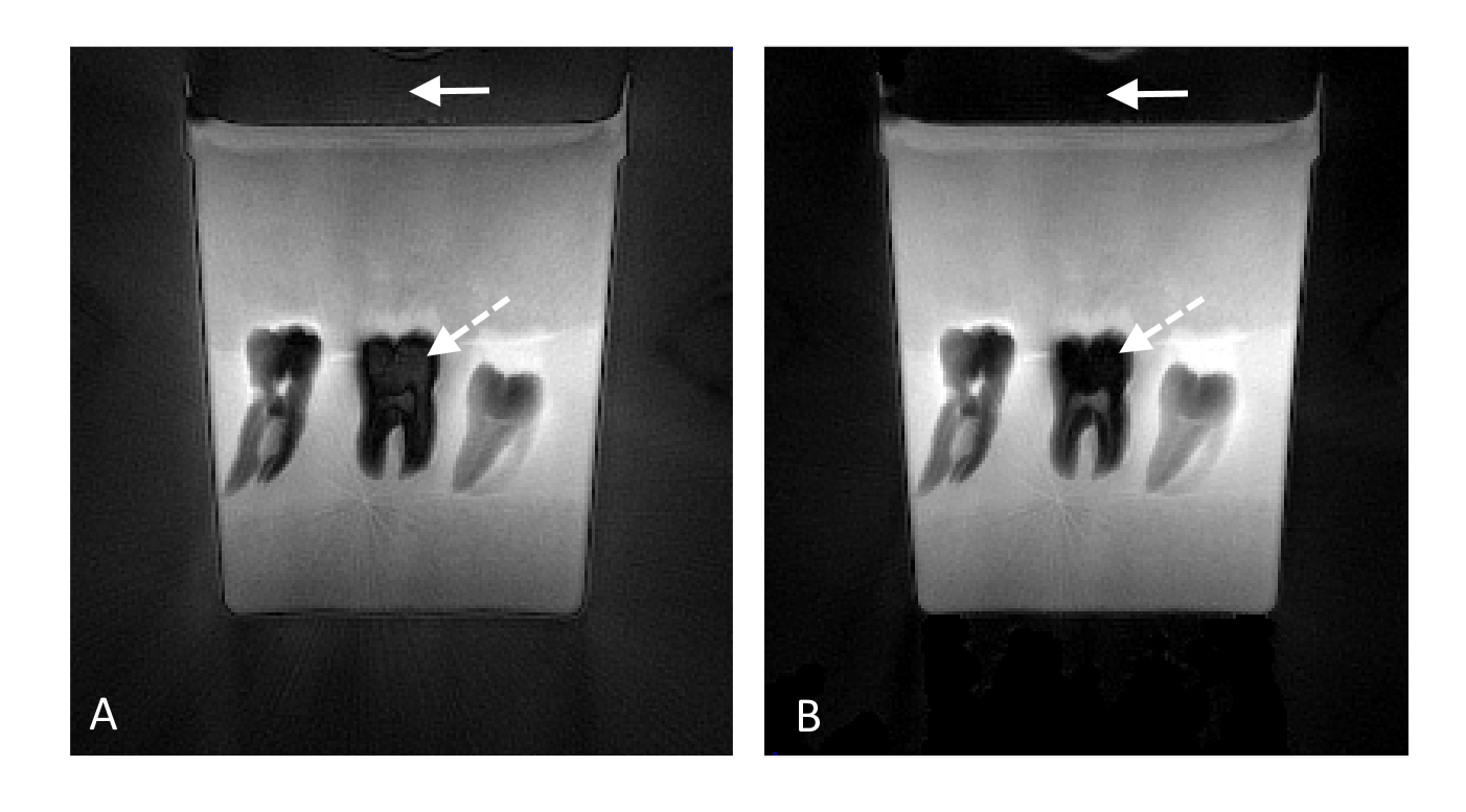
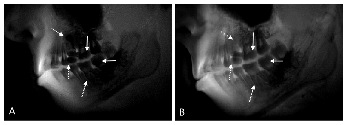
Figure 2 demonstrates the well-defined slice selectivity of the modified 2D-UTE sequence. Removal of corrupted data points and compressed sensing based reconstruction successfully corrected the DC-offset caused by the transient phase of the FIR filter and yet detuned coil (Figure 3). Figure 4 shows acquired MR pantomograms at 1.5T field strength with a standard 2D-UTE, TE=750µs (image A) and the 240µs half-pulse acquisition, TE=35µs (image B). The lower echo time and reduced signal decay with 240µs pulses led to the detection of signal in dentin and show the roots to be clearly distinguishable from the periodontium. The extremely short echo time also allowed for less dephasing due to susceptibility changes and reduced the artifacts caused by metal implants.Conclusion
The presented modified 2D-UTE sequence has been successfully applied to in vivo acquisition of MR pantomograms with signal detection from hard dental tissue at 1.5T field strength. In combination with the quadratic-phase saturation pulses an excellent slice profile could be obtained. It provides an effective alternative to 3D-UTE methods in cases isotropic spatial resolution is not required and a radiation free alternative to the standard x-ray based pantomograms.Acknowledgements
No acknowledgement found.References
[1] Pauly J, Nishimura D, Macovski A. A k-space analysis of small-tip-angle excitation. J Magn Reson 1989;81:43–56
[2] Conolly S, Nishimura DG, Macovski A, Glover G. Variable-rate selective excitation. J Magn Reson 1988;78:440–458
[3] Josan S, Kaye E., Pauly J, Daniel B, Butts Pauly K. Improved Half RF Slice Selectivity in the Presence of Eddy Currents with Out-of-Slice Saturation. Magn Reson Med 2009;61:1083–1089
[4] Lustig M, Donoho D, Santos J, Pauly J. Compressed sensing MRI. IEEE Signal Process. Mag. 2008;25(2):72–82
Figures