4025
Whole-Body, Zero TE Based Pseudo CT Conversion1GE Global Research, Munich, Germany, 2GE Global Research, Schenectady, NY, United States, 3GE Global Research, Bangalore, India, 4Radiology and Biomedical Imaging, UCSF, San Francisco, CA, United States
Synopsis
Proton density (PD) weighted Zero TE (ZTE) MR imaging has been demonstrated to provide accurate bone depiction, segmentation and pseudo CT conversion in the head. However, when applied for the whole-body, discriminating between air and bone appears challenging (primarily because of SNR and RF shading). Here we present a novel method for decomposition of low ZTE signal intensity regions into bone and air based on connected component and shape analysis. The method is demonstrated for whole-body, pseudo CT conversion in three PET/MR patients.
Purpose:
Proton density (PD) weighted Zero TE (ZTE) MR imaging has been demonstrated to provide accurate bone depiction, segmentation and pseudo CT conversion in the head [1,2]. However, when applied for the whole-body, discriminating between air and bone appears challenging (primarily because of SNR and RF shading). Here we present a novel method for decomposition of low ZTE signal intensity regions into bone and air based on connected component and shape analysis. The method is demonstrated for whole-body, pseudo CT conversion in three PET/MR patients.Methods:
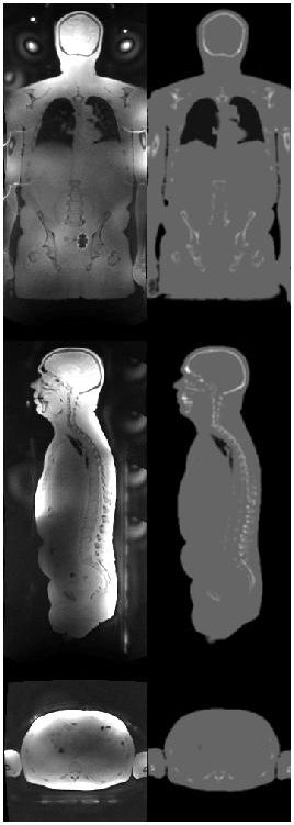
Whole-body, zero TE imaging was performed using PD-weighted parameter settings: FA=1deg, BW=250kHz, FOV=500mm, res=2.6mm, number of 3D radial spokes = 110592, scan time 1min24sec per bed. The method was first tested in volunteers using a GE 3T MR750w scanner, followed by three patient scans using a 3T, time-of-flight (TOF) GE Signa PET/MR scanner and GEM whole-body, surface coil receive (GE Healthcare, Waukesha, WI). Scanner reconstructed DICOM images from the individual bed positions were stitched and bias corrected via low-pass filtering (cf. Fig. 3: left). The body contour was segmented and a low ZTE signal mask (MASKlowZTE) inside the body was detected using thresholding of the original and the bias-corrected ZTE images. The obtained MASKlowZTE was decomposed into internal air and bone components using connected component analysis (CCA). A whole-body bone skeleton (BONE) was generated by selectively adding the bone components from the 12 largest connected components found. Pseudo CT conversion was done by assigning fixed Hounsfield units for air (-1000HU) and soft-tissue (0HU), while using a linear signal scaling for the bone mask, according to: pCT(BONE) = -2000*(ZTE(BONE)-1) [2,3].Results:
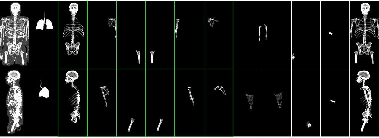
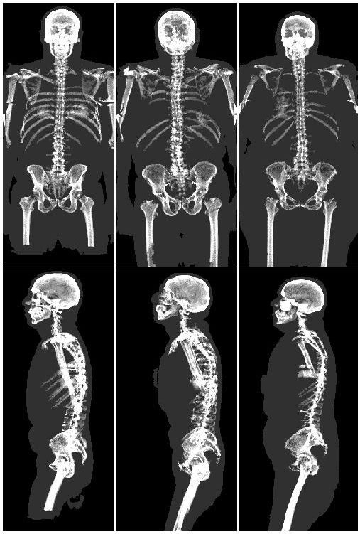
Figure 1 illustrates whole-body ZTE bone decomposition for one representative patient in coronal (top) and sagittal (bottom) projections. The left image shows the projected low ZTE signal mask MASKlowZTE on top of the body contour (gray), including both bone and internal air regions. The images in the middle, show the 12 largest connected components depicting the major bones (skull, pelvis, vertebra, ribs, femur, humerus, shoulders, clavicle …) and air regions (lungs, abdominal gas, air layers sandwiched in between body parts, ...). The image on the right shows the selective superposition of the bone components (BONE) clean of internal air. Figure 2 summarizes the results for all three patients and Figure 3 illustrates the converted pseudo CT along three orthogonal slices (right) next to the stitched, whole-body ZTE (left).Discussion:
Proton density weighted ZTE imaging is ideally suited for whole-body bone, tissue, air segmentation. More specifically, zero TE images demonstrate uniform soft tissue signal response without T1 saturation (FA=1deg), or destructive signal interference at fat/water tissue interfaces (TE=0 and BW=250kHz). Low ZTE signal regions corresponding to either bone or internal air, can effectively be separated based on connected component analysis. Selectively adding the bone components (from the 12 largest connected air/bone regions) depicts all major skeletal bone structures clean of remaining air artifacts. Differentiation between air and bone connected components can be automated using e.g. anatomical prior knowledge, shape properties, or other information like Joint Estimation in a PET/MR environment [4, 5]. The described methods can be used for several applications, including: PET/MR attenuation correction, MR-based Radiation Therapy Planning (RTP), and musculoskeletal MR imaging.Acknowledgements
No acknowledgement found.References
[1] Wiesinger et al, Zero TE MR bone imaging in
the head, MRM 75(1): 107 (2016).
[2] Wiesinger et al, Whole Body Skeletal Imaging Using Zero TE, ISMRM
Singapore, p.675 (2016).
[3] Leynes et al, Quantitative Evaluation of the Effect of Bone on Pelvic Lesion Uptake for MR-Based Attenuation Correction on an Integrated Time-Of-Flight PET/MRI System, ISMRM Singapore, p.2451 (2016).[4] Defrise et al, Time-of-flight PET data determine the attenuation sinogram
up to a constant, PMB 57(4): 885 (2010)
[5] Ahn et al, Robust PET Attenuation Correction for PET/MR Using Joint
Estimation with MR-Based Priors, ISMRM Singapore, p. 1776 (2016).
Figures