4011
Ultrashort echo time imaging at 1.5 T using an insertable unshielded gradient coil and cone trajectoriesAyana Setoi1 and Katsumi Kose1
1University of Tsukuba, Tsukuba, Japan
Synopsis
Ultrashort echo time (UTE) imaging with cone trajectories was installed to a 1.5T compact MRI system using an unshielded insertable gradient coil. K-trajectories of the 3D cone trajectory acquisition were measured using a small capillary phantom and used for image reconstruction. A LEGO block sample with T2* of about 0.6 ms was successfully imaged with echo time of 0.05 ms to 0.6 ms. This result demonstrated that UTE imaging sequences with cone trajectories were successfully installed to our system.
Introduction
Ultrashort echo time (UTE) imaging is widely used for MRI of T2 or T2* short materials such as cortical bones, tendons, and lung tissues. Because UTE imaging requires short and intense RF pulses, and fast and intense field gradients, its implementation to existing whole body scanners has several difficulties. In this study, we solved these problems using an insertable gradient coil and cone trajectories with k-space trajectory correction.Materials and methods
We used a home-built compact MRI system consisting of a 1.5 T and 280 mm diameter horizontal bore superconducting magnet (JMBT-1.5/280/SS, JASTEC, Kobe, Japan), a second-order room temperature shim coil set (diameter = 135 mm), an unshielded gradient coil set designed with the target field method [1] (diameter = 105 mm, efficiencies: 4.48, 4.73, and 4.95 mT/m/A for Gx, Gy, and Gz), an eight-rung birdcage coil (diameter = 64 mm, length = 64 mm), and a digital MRI console (MRTechnology, Tsukuba, Japan). Figure 1 shows samples used for UTE imaging and k-space trajectory correction. Cone trajectories used for 3D image acquisition were developed by combining spiral trajectories designed by the Glover’s approach [2] and a radial sampling along the other direction (e.g. z direction). The polar angle from z direction (0~π) was equally divided into 128. The number of shots for one cone was 8, 16, 32, and 64. K-space trajectories were measured using a water phantom in a glass capillary (inner diameter = 1.0 mm, water length = 15mm). The k-trajectories were measured in the xy-plane and in the xz plane by placing the capillary phantom along z and y directions, respectively. The trajectories were calculated from the phase of the NMR signal of the capillary phantom. The SNR of the NMR signal was improved by 1,024 times signal accumulation. UTE images of the samples were acquired with cone trajectories some delay times after a 60° RF pulse (40 µs width) or a 90° RF pulse (120 µs width). The repetition times of the sequences were 400 ms.Results
Figures 2(a) and (b) show temporal changes of (a) kx and ky and (b) kx and kz, and k-trajectories in (a) xy and (b) xz plane measured using the capillary phantom. When the time t is smaller than about 0.5 ms, considerable distortion of the k-trajectories was observed. Figure 3 shows 2D cross-sections selected from 3D image datasets of the cylindrical phantom acquired with the cone-trajectories. The field of view was 64 mm cube and image matrix was 128×128×128. Figures 3(a) and (b) were acquired when the cylindrical phantom was placed parallel to the xy plane and parallel to the xz plane and reconstructed using theoretical and measured trajectories. Figures 4(a) and (b) show 2D cross-sections selected from 3D image datasets of the LEGO block sample acquired with (a) 256-shot radial trajectories for one polar angle and (b) 64-shot cone trajectories for one polar angle. The FOV and image matrix were identical to those of Fig.3. The spatial resolution is comparable between (a) and (b) but the acquisition time of (b) was about 1/4 of that of (a) except the overhead time of the sequence updates. Figure 5 shows 2D cross-sections selected from 3D image datasets of the LEGO block sample acquired with cone trajectories when (a) a 120 µs width square 90° RF excitation pulse and (b) a 40 µs width square 60° RF excitation pulse were applied. The image intensity of the LEGO block sample monotonously decayed with the echo time, which reflected T2* (~0.6ms) of the sample.Discussion
As shown in Fig.2, considerable distortion was observed for the k-trajectories when the time t was smaller than 0.5 ms. We think that this distortion was caused by contamination from NMR signals of solid-like materials such as epoxy resin to seal the water in the capillary and some materials used to fix the capillary phantom. This distortion was not harmful for k-trajectory correction because the theoretical trajectories can be used for image reconstruction after overall fitting of the trajectories. Figure 3 demonstrates that k-trajectory correction worked well. Figure 4 shows the 3D UTE imaging using cone trajectories (64 shots for one cone angle) was three to four times faster than that using radial trajectories with similar spatial resolution. Because the T2* of the LEGO block protons were about 0.6 ms, Figure 5 shows the 3D UTE imaging using cone trajectories were successfully installed to our system. In conclusion, 3D UTE imaging using an insertable gradient coil can be a useful approach to 3D MRI of solid-like materials in whole body MRI systems.Acknowledgements
No acknowledgement found.References
[1] Turner R. A target field approach to optimal coil design. J. Phys. 1986; D19: L147-151
[2] Glover G H. Simple analytic spiral k-space algorithm. Magn Reson Med 1999; 42: 412.
Figures
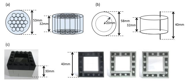
(a) Cylindrical water phantom consisting of 19 acrylic pipes (8 mm
diameter, 1 mm thick) filled with CuSO4 water solution. (b)
Capillary phantom fixed in an acrylic cylinder used for k-trajectory
measurements. The inner diameter was 1.0 mm and length of water was about 15
mm. (c) LEGO block sample.
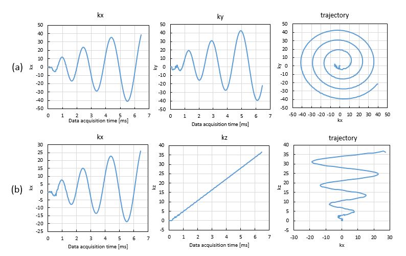
(a) Temporal change of kx
and ky and the k-trajectory measured in the xy-plane for 16-shot sequence.
(b) Temporal change of kx and kz and the k-trajectory
measured in the xz-plane for 16-shot sequence.
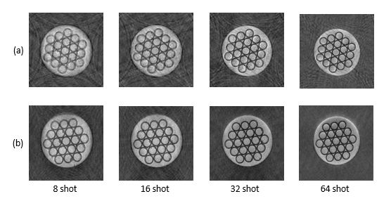
2D cross-sections selected from 3D image datasets acquired with cone-trajectories
(TR=400ms, TE=0.6ms). (a) The phantom was placed parallel to the xy plane. (b) The
phantom was placed parallel to the xz plane. These images were reconstructed
with theoretical and measured k-trajectories.
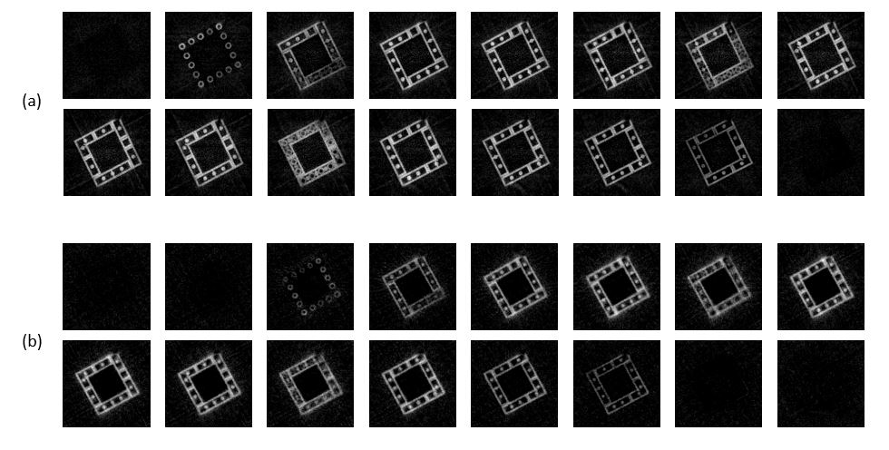
2D cross-sections selected from 3D image datasets of the LEGO block sample.
Displayed in 1 mm intervals. (a) 256-shot radial trajectories for one polar
angle. (b) 64-shot cone trajectories for one polar angle. The spatial
resolution was comparable but the acquisition time of (b) was about 1/4~1/3 of
that of (a).
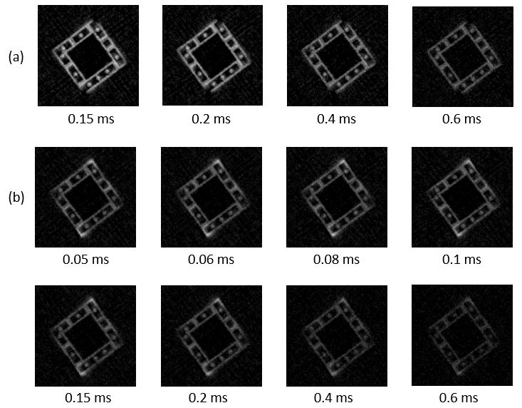
2D cross-sections selected from 3D image datasets of the LEGO block
sample acquired with cone trajectories with various delay time after the RF
pulse. (a) Acquired with a 120 μs width
square 90° RF
pulse. (b) Acquired with a 40 μs
width square 60° RF
pulse.