3983
Boosting SNR and/or Resolution of Arterial Spin Label (ASL) imaging using Multi-contrast Approaches with Multi-lateral Guided Filter and Deep Networks1Electrical Engineering, Stanford University, Stanford, CA, United States, 2Radiology, Stanford University, Stanford, CA, United States
Synopsis
Arterial Spin Labeling (ASL) MRI is a powerful neuro imaging tool which provides quantitative perfusion maps. However, ASL perfusion maps typically suffer from low SNR and resolution. Averaging from multiple scans (high Nex value) can improve the SNR but at the cost of significantly increased acquisition time. In the work we proposed a technique for improved ASL image quality with boosted SNR and/or resolution by 1) incorporating the information of multi-contrast images 2) using nonlinear, non-local, spatial variant multi-lateral filtering, 3) training a deep network model to adaptively tune the final denoising level and further boost the SNR and improve image quality. Various in-vivo experiments demonstrate the superior performance of the proposed method which will significantly accelerate ASL acquisition and improve image quality.
Introduction
Arterial Spin Labeling (ASL) MRI uses the signal differences between labeled and control image to quantify the blood perfusion. It is a powerful MRI technique and is applied increasingly for both research studies and clinically for neurological, cerebrovascular and psychiatric diseases. However, ASL perfusion maps typically suffer from low SNR due to its signal subtraction. The SNR can be increased in the order of square-root of $$${Nex}$$$ if the ASL scans are repeated $$${Nex}$$$ times. 3 or even more repetition times are applied typically to achieve an acceptable image quality. However repeating the scans significantly increase the scan duration and is therefore not feasible for certain applications, like acute stroke etc. Recently proposed multi-delay ASL [1] can compensate the effect of various transit delays for better sensitivity of perfusion measurement and the measurement of arterial transit time (ATT). However, acquiring multiple delays further increases the time cost and results in even lower SNR and resolution due to the time constraints. Here we proposed a solution to this urgent issue of SNR starvation in ASL imaging that will significantly improve image quality per unit time.Methods
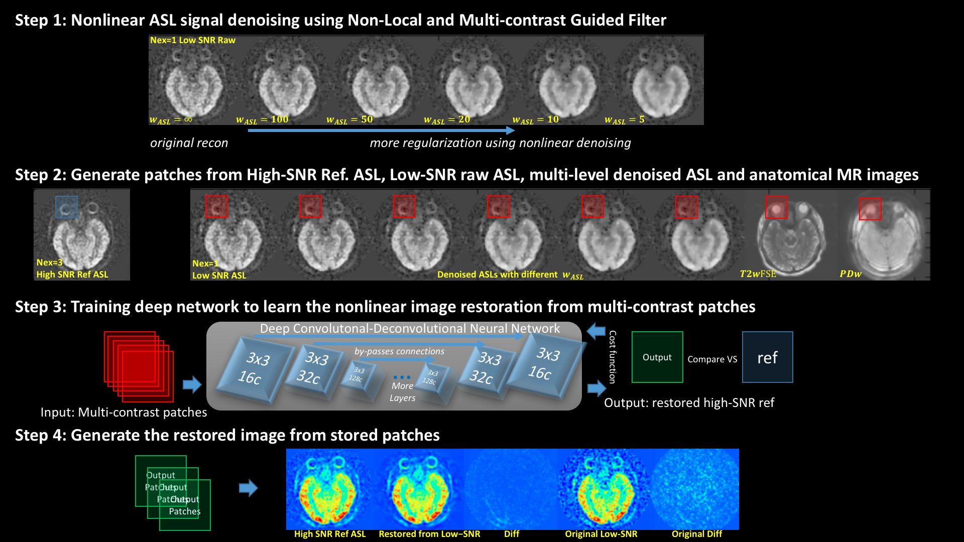
There are 3 main innovative contributions of this proposed work: 1) Incorporation of multi-contrast information in ASL denoising 2) Application of a nonlinear spatial-variant filter to prevent over-smoothing of edges, and 3) Generation of the final denoising/restoration result using a deep network.
The workflow of the proposed method is shown in figure1.
1. Multi-lateral guided filtering using multi-contrast information. For ASL exam there is typically one PDw image taken without labeling. There are also highly likely to be additional anatomical scans, such as T1w, T2w, and FLAIR, acquired as part of any clinical protocol. These images share the basic structure information and have much higher SNR. Here we used a Multi-lateral Guided Filter [2] to conduct location-variant weighted average for each pixel. Derived from the bilateral filter, the Multi-lateral filter assigns weight $$$g(I(x),I(\xi))$$$ as a product of the square-differences of ASL signal and multi-contrast anatomy MR (T2w, PDw, etc.) signals from each pixel $$$x$$$ to its neighbor (5x5) pixel $$$\xi$$$. Different from conventional Gaussian or Wavelet based [3] denoising, this step is a nonlinear filter, which tends to better preserve structures and avoid over-smoothing. Weighting parameter $$$w_{ASL}$$$ here controls the denoising strength.
Bilateral Filter:
$$Bilateral(x)=\frac{\sum_\xi{k(x,\xi)g(I(x),I(\xi))I(\xi)}}{\sum_\xi{k(x,\xi)g(I(x),I(\xi))}}$$
Extend to Multi-lateral Filter as Bilateral filter in a higher dimensional space:
$$k(x,\xi)=\exp( {-\frac{(x-\xi)^2}{8}})$$
$$I(\xi)=I_{ASL}(\xi)$$
$$g(I(x),I(\xi))=g_{ASL}(I_{ASL}(x),I_{ASL}(\xi))g_{T2w}(I_{T2w}(x),I_{T2w}(\xi))g_{PDw}(I_{PDw}(x),I_{PDw}(\xi))$$
$$g_{T2w}(a,b)=g_{PDw}(a,b)=\exp( {-\frac{(a-b)^2}{2}})$$
$$g_{ASL}(a,b)=\exp{(-w_{ASL}(a-b)^2)})$$
2. Forming ASL+multi-contrast MRI patches. After the denoising, we reconstructed multiple versions denoised ASL with different $$$w_{ASL}$$$. A stack of multi-contrast images were formed including: the original low-SNR ASL, multiple denoised ASL with different smoothing levels, co-registered T2w image and the unlabeled PDw image. We cropped small multi-contrast patches (16x16 etc.) from these multi-contrast images. The final denoising employs these local stacks of patches which accelerates computation, reduces model complexity and increases the training sample size (~10000 patches from 1 single slice) to prevent any overfitting in the deep network training.
3. Deep Network for Denoising Reconstruction. We then trained a deep network to output the final denoising and restoration results. Here we used a convolutional-deconvolutional neural network [4] with the structure as the image shows. The input of the deep network is the multi-contrast MRI patches, and the output is the final denoised version. The deep network is trained on one set of slices using high-SNR/high-resolution ASL as ground-truth and applied on different scans and slices.
4. The final denoised ASL is formed by synthesizing the output patches.
Results
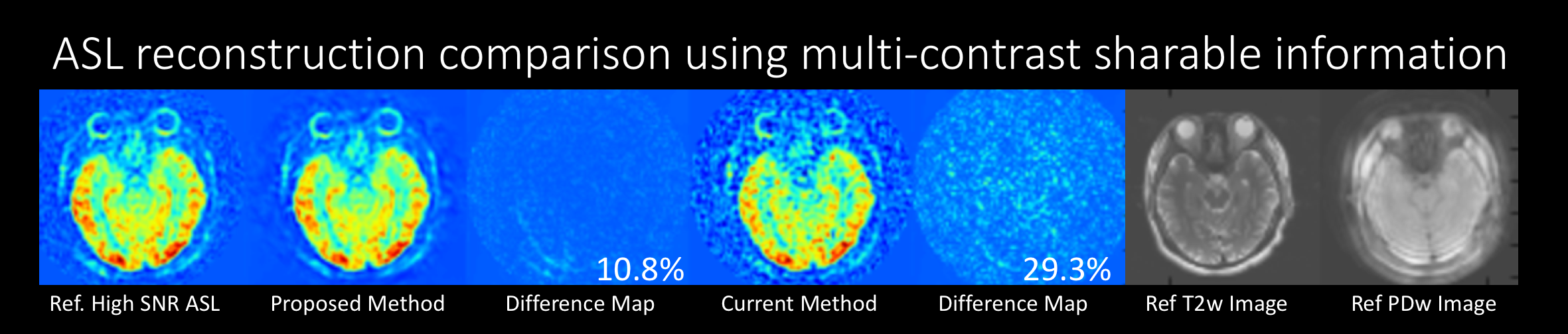
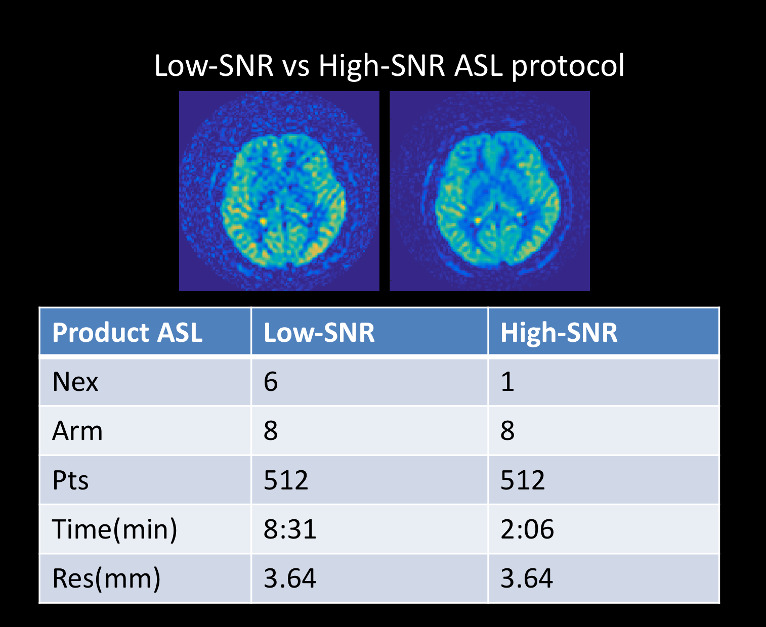
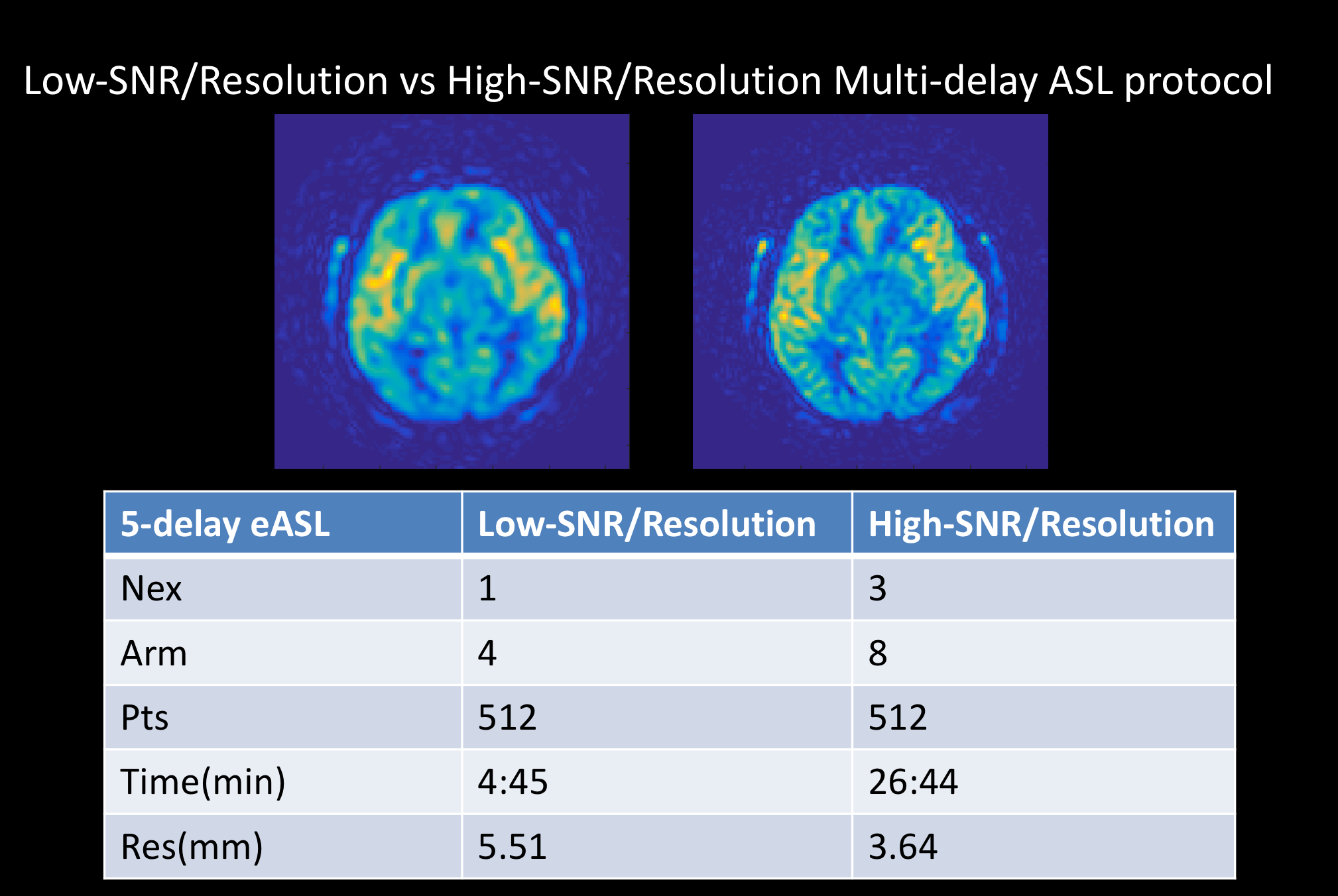
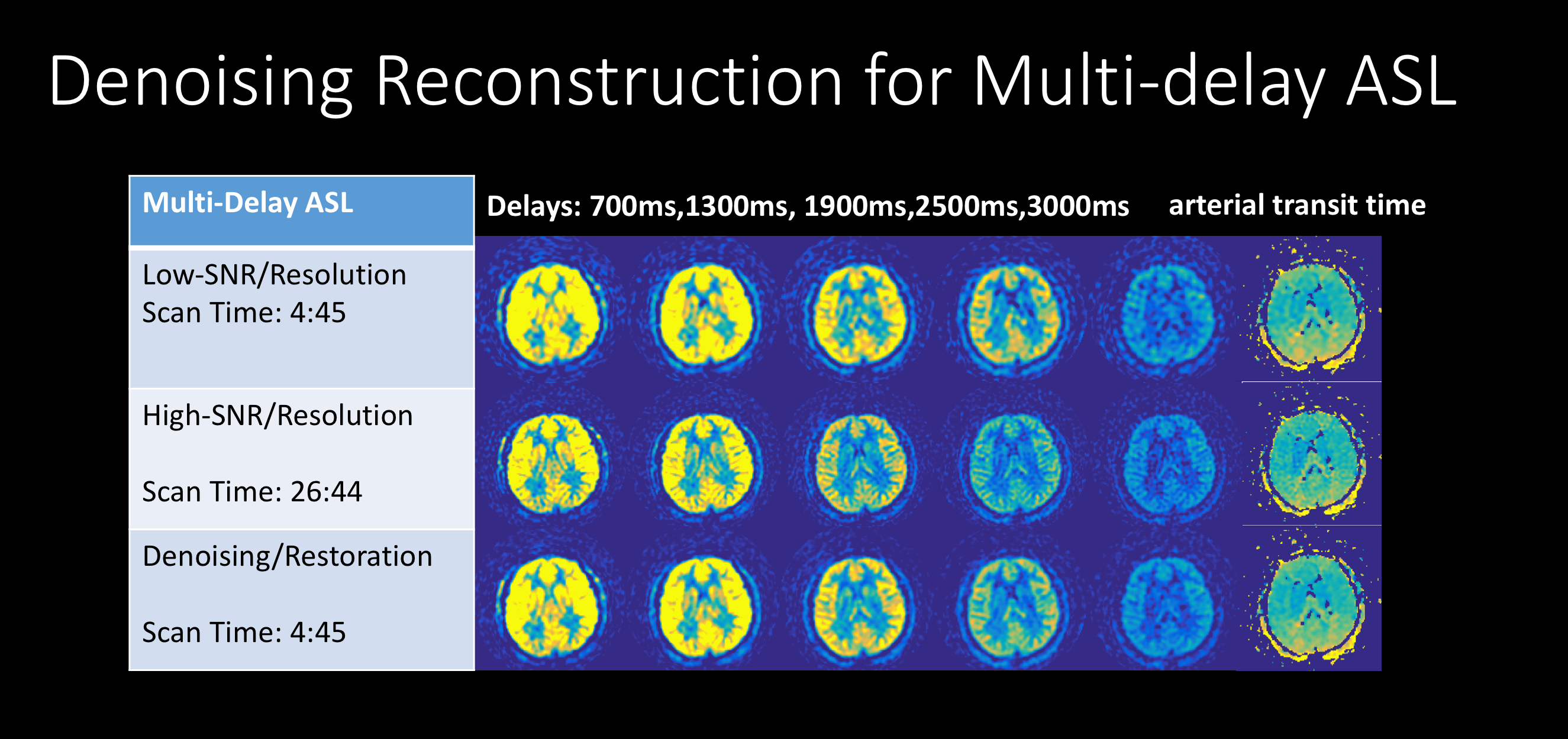
Here we conducted multiple in-vivo experiments. First we validated the performance of the proposed algorithm for improving SNR. Using Nex=6 as reference high-SNR ASL, the results shown the proposed algorithm can reduce the error and noise for low-SNR ASL (Nex=1), 4 times shorter absolute acquisition time compared to the high-SNR-ASL reference scan. Then we validated the performance of the proposed algorithm on improving both SNR and resolution in multi-delay ASL(700ms,1300ms, 1900ms,2500ms,3000ms). The results demonstrate better image quality for each delay as well as improved arterial transit time mapping [5].Discussion and Conclusion
In-vivo experiments demonstrate the proposed method provides superior performance for restoring ASL image with higher SNR and/or resolution (effectively as ~6+Nex or 4+time reduction). By incorporating multi-contrast information, using Multi-lateral Guided Filter and Deep Networks, the proposed method out-perform existing reconstruction and denoising results can better reduce noise, preserve structures and provide more detailed functional metrics such as CBF and transit-time maps. The proposed algorithm can also be applied to complement Parallel Imaging and Compressed Sensing for further acceleration of ASL scans.Acknowledgements
Here we acknowledge GE Healthcare for supporting this work.References
[1] Dai, Weiying, et al. "Reduced resolution transit delay prescan for quantitative continuous arterial spin labeling perfusion imaging." Magnetic resonance in medicine 67.5 (2012): 1252-1265.
[2] He, Kaiming, Jian Sun, and Xiaoou Tang. "Guided image filtering." European conference on computer vision. Springer Berlin Heidelberg, 2010.
[3] Bibic, Adnan, et al. "Denoising of arterial spin labeling data: wavelet-domain filtering compared with Gaussian smoothing." Magnetic Resonance Materials in Physics, Biology and Medicine 23.3 (2010): 125-137.
[4] Xie, Junyuan, Linli Xu, and Enhong Chen. "Image denoising and inpainting with deep neural networks." Advances in Neural Information Processing Systems. 2012.
[5] Dai, Weiying, et al. "Reduced resolution transit delay prescan for quantitative continuous arterial spin labeling perfusion imaging." Magnetic resonance in medicine 67.5 (2012): 1252-1265.
Figures