3982
Artificial Neural Network for Suppression of Banding Artifacts in Balanced Steady-State Free Precession MRI1Department of Bio and Brain Engineering, Korea Advanced Institute of Science and Technology (KAIST), Daejeon, Korea, Republic of, 2Graduate School of Medical Science and Engineering, Korea Advanced Institute of Science and Technology (KAIST), Daejeon, Korea, Republic of
Synopsis
This study is the first attempt for a learning-based algorithm to be applied to banding artifact suppression in balanced steady-state free precession (bSSFP). We trained multilayer perceptron (MLP) models with two or four phase‑cycling datasets and banding-free datasets as inputs and outputs, respectively. We demonstrated that MLP was superior to existing methods in terms of banding artifact suppression and SNR efficiency, which was clearer in two phase‑cycling datasets. Furthermore, MLP was widely applicable to various image sets, irrespective of scan parameters, body organs, and field strengths. The learning-based approach is promising for banding artifact suppression of bSSFP.
Introduction
Balanced steady state free precession (bSSFP) sequence has been gaining importance in clinical practices due to its high speed, high signal-to-noise ratio (SNR), and minimal distortion. However, bSSFP often suffers from well-known banding artifacts, which are related to the field strength and repetition time (TR) (1). Various methods have been introduced to suppress banding artifacts in bSSFP, but banding artifacts cannot be suppressed completely by these methods, especially when number of multiple phase cycling (PC) bSSFP datasets is small (2-4). In this study, we proposed a learning-based method to combine the bSSFP datasets with multiple PC for better banding artifact suppression. Models of multilayer perceptron (MLP), a feedforward artificial neural network (ANN), were trained to predict banding free bSSFP images based on two and four PC datasets. Then, the trained MLP models were applied to another bSSFP datasets for evalulation in terms of performance of banding artifact suppression.Materials and Methods
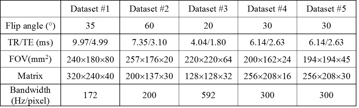
MLP consists of three layers of input, hidden, and output layers. In training stage, inputs and outputs of the MLP models were two or four PC datasets and banding-free images, respectively. Banding-free bSSFP images were generated by maximum-intensity projection (MIP) of 8 or 12 phase-cycled datasets. In test stage, the learned MLP models received two (0 and 180°) or four (0, 90, 180, and 270°) PC datasets as input and produced banding-suppressed images as output. The training and test were performed in two ways. First, Bloch equation simulations were performed to generate 12 PC datasets. The ground truth was reconstructed by MIP of all PC datasets, among which two (0 and 180°) PC datasets were used as the input. The MLP models were trained by the inputs and the ground truth, and the trained models were evaluated with simulated ‘bi-tissue’ datasets with T1/T2= 420/50 ms and 1100/70 ms. Two separate 3D bSSFP datasets with multiple PCs were acquired in humans; axial brain bSSFP images (dataset #1) in one subject (subject #1) using 8 PCs and coronal knee bSSFP images (dataset #2) in another subject (subject #2) with 12 PCs. Using all PC datasets, the ground truth images and the inputs were produced in the same way as the simulation. To test the trained MLP models, axial brain images (dataset #3), sagittal knee images (dataset #4), and axial knee images (dataset #5) were acquired. The MLP models were trained by two or four PC datasets as input and were additionally evaluated in human experiments. Image parameters of all the scans were shown in Table 1. The mean square error (MSE) index was used to measure the difference between the ground truth images and the outputs of MLP, MIP, and sum-of-square (SOS), and SNR was also calculated. All experiments were performed with a 3T whole-body scanner (Siemens Medical Solutions, Erlangen, Germany). Image analysis was performed using MATLAB(The Mathworks, Natick, MA).Results
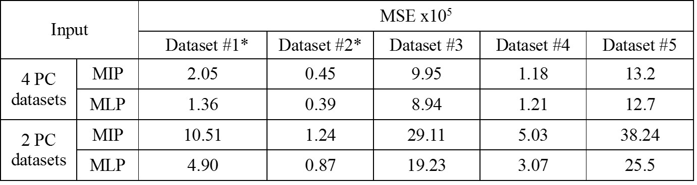
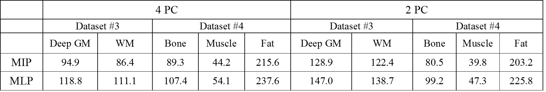
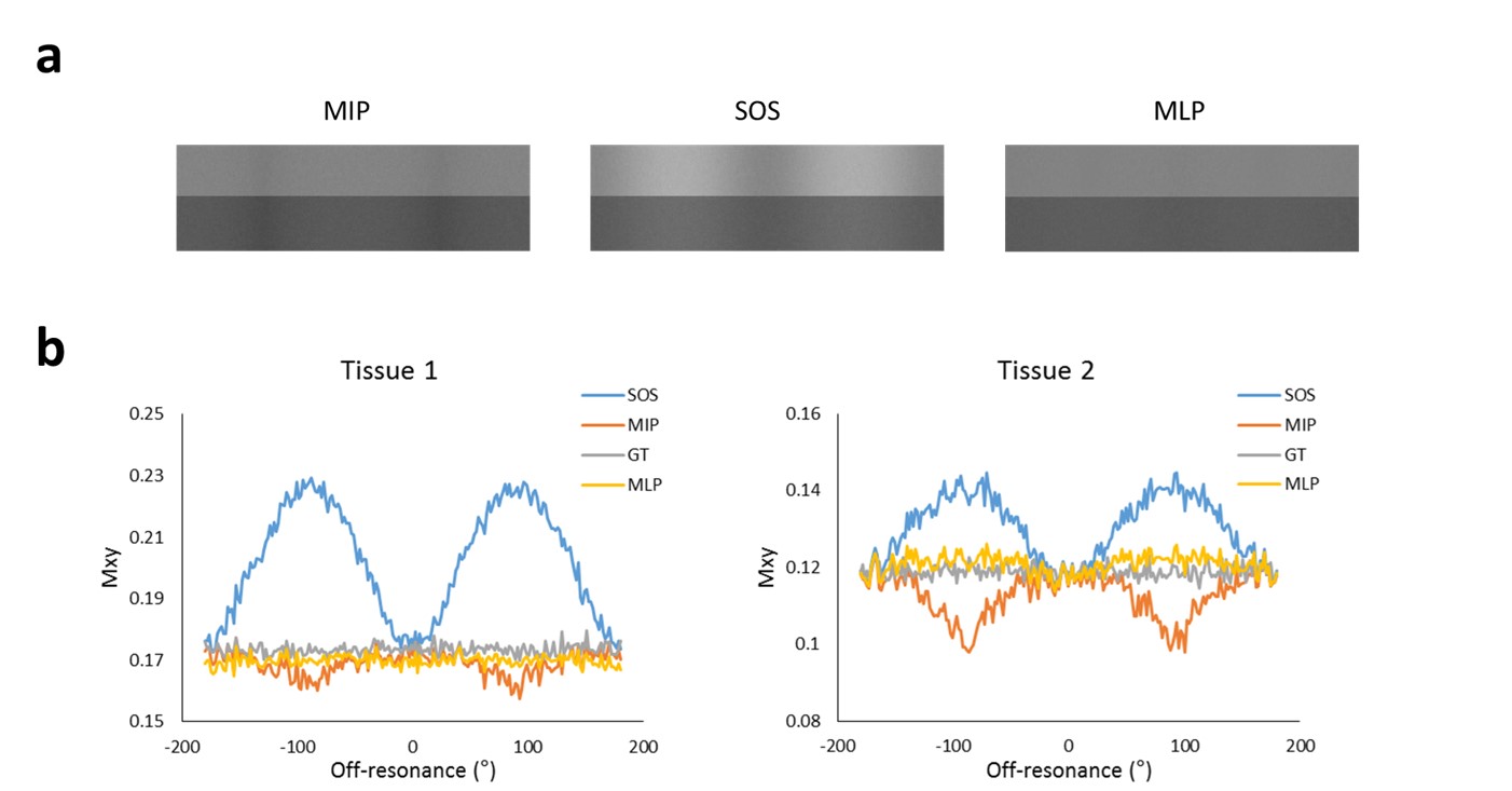
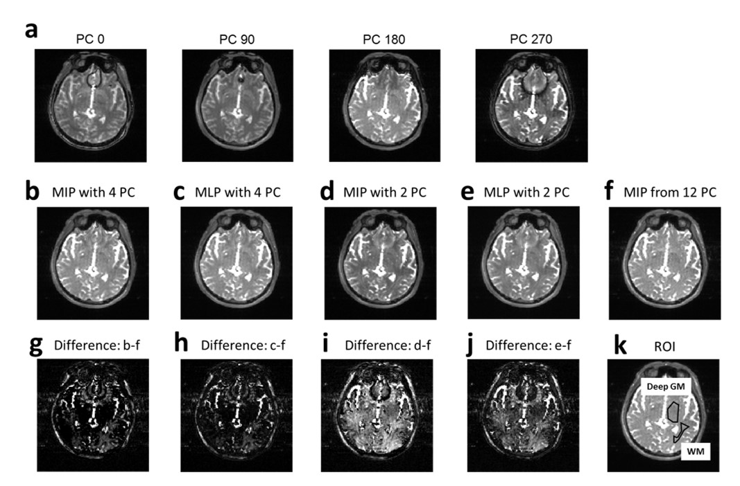
The simulation showed that MLP was superior to the existing methods (MIP and SOS) for banding artifacts suppression (Figure 1): The images combined with MIP and SOS demonstrated visible signal fluctuations, which were mostly eliminated in the results of MLP. MSE values of MLP (1.5X10⁻⁵) were much smaller than those of the existing methods (MIP: 5.6x10⁻⁵, SOS: 6.7x10⁻⁴). The results of human experiments were consistent with those of simulations. MLP showed lower MSE values than those of MIP in both training datasets and test datasets (Table 2), except for the four PC images of dataset #3. The bSSFP images at different PC angles showed banding artifacts at different spatial locations (Figure 2a). Visually both MIP and MLP suppressed banding artifacts well (Figure 2b-e), however, the differences between the ground truth images and MIP/MLP images were clearly visualized in the subtraction images (Figure 2g-j). MLP showed smaller difference than that of MIP. MLP showed higher SNR efficiency than MIP in all tissues (Table 3).Discussion and Conclusion
To our knowledge, it is the first time for a learning-based algorithm to be applied to reducing banding artifacts in bSSFP. Combination of multiple PC datasets with MLP successfully suppressed banding artifacts in bSSFP MRI, while maintaining high SNR. The MLP models trained with a couple of 3D multiple PC datasets from two different organs in human worked well on datasets acquired with different scan parameters from different subjects, and also those acquired from a different species at different field strength. MLP worked well even with two PC datasets, providing better banding artifact suppression and higher SNR efficiency than the widely-used MIP. Artificial neural network is a promising approach to combining multiple phase cycled bSSFP datasets for banding artifact suppression.Acknowledgements
No acknowledgement found.References
1. Park SH, Han PK, Choi SH. Physiological and Functional Magnetic Resonance Imaging Using Balanced Steady-state Free Precession. Korean journal of radiology 2015;16(3):550-559.
2. Haacke EM, Wielopolski PA, Tkach JA, Modic MT. Steady-state free precession imaging in the presence of motion: application for improved visualization of the cerebrospinal fluid. Radiology 1990;175(2):545-552.
3. Vasanawala SS, Pauly JM, Nishimura DG. Linear combination steady-state free precession MRI. Magnetic resonance in medicine 2000;43(1):82-90.
4. Bangerter NK, Hargreaves BA, Vasanawala SS, Pauly JM, Gold GE, Nishimura DG. Analysis of multiple-acquisition SSFP. Magnetic resonance in medicine 2004;51(5):1038-1047.
Figures