3973
B0 Mapping of Highly Inhomogeneous Fields using Missing-Pulse Steady-State Free Precession (MP-SSFP)1Center for Magnetic Resonance Research,Department of Radiology, University of Minnesota, Minneapolis, MN, United States
Synopsis
A B0 mapping of highly inhomogeneous fields is introduced. The method acquires refocused echo signals generated by a missing-pulse steady-state free precession (MP-SSFP) sequence with three-dimensional phase-encoding gradients. A B0 map is calculated from the refocusing and dephasing of spin phase around the echo centers. Validation was performed in a phantom experiment conducted with a permanently inhomogeneous field produced by mounting a head gradient coil at 36 cm out of the isocenter of a 90-cm 4T magnet. Using the measured B0 field map, we demonstrate correction of image distortion caused by the extremely nonlinear inhomogeneous B0 field.
Purpose
Increased access to MRI scanning can occur if the cost of the MRI system can substantially decrease. One approach to achieve this is to decrease the size of MRI magnets, but this comes at a cost of drastically reduced static field (B0) homogeneity, which can easily lead to >100 kHz off-resonance variation. Therefore, MRI methods that tolerate extreme B0 inhomogeneity are needed. Here we introduce a method to map the B0 of a highly inhomogeneous magnet, far exceeding capabilities of conventional approaches (e.g., using two gradient echo images with different echo times). The method is a variant of the missing pulse steady-state free precession (MP-SSFP) sequence1 that works under strong permanent B0 inhomogeneity.Methods
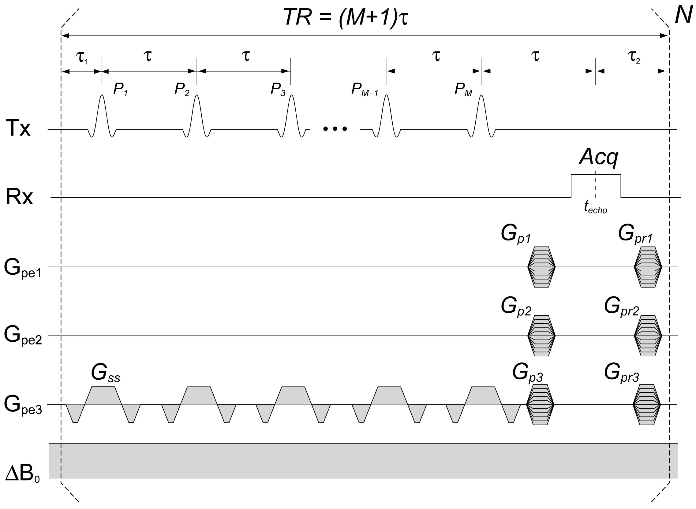
A permanently inhomogeneous B0 was introduced by mounting a head gradient coil at 36 cm away from the magnet center of a 4T human MRI system (Agilent technologies, inc.). With the MP-SSFP sequence, echoes consisting of overlapping spin and stimulated echoes refocus at the missing pulse timings regardless of the field inhomogeneity (Fig.1); spin isochromats refocus toward the echo peak timing, techo, and then dephase after the echo peak. Spatial encoding was performed with three phase encoding gradients and data were sampled around the refocusing echo peak. Thus, reconstructed images were 4-dimensional (3 spatial and 1 time dimensions). A 3D B0 map [Hz] was calculated from images at two time points around techo (I1 and I2 for t1 and t2) by $$B_{0}=\frac{arg(I_{2})-arg(I_{1})}{2\pi(t_{2}-t_{1})}$$
Under the inhomogeneous field, B0 mapping using a cylindrical phantom filled with distilled water and cubic Teflon objects inside was performed with the following sequence parameters: τ = 2.4 ms, pulse missing is every 10 pulses (TR = 24 ms), sinc pulse excitation with FA = 2°, BW = 200 kHz and Tp = 30 μs (time-bandwidth product TBP = 6), readout bandwidth = 250 kHz, FOV = 20x20x20 cm, matrix size = 48x48x64 and scan time = 59 min. A quadrature TEM coil was used for transmit and receive. 3D MP-SSFP measurement using the inhomogeneous field gradient for spatial encoding was also performed. Since the inhomogeneous field gradient was not linear, an image from 3D MP-SSFP showed image distortion. The image distortion was corrected by calculating an image distortion field based on the measured B0 map. Signal intensities were also corrected by the Jacobian of the image distortion field. Image reconstruction was performed with a reconstruction routine written with C++ and image distortion and intensity correction were done with Insight Toolkit (ITK)2.
Results
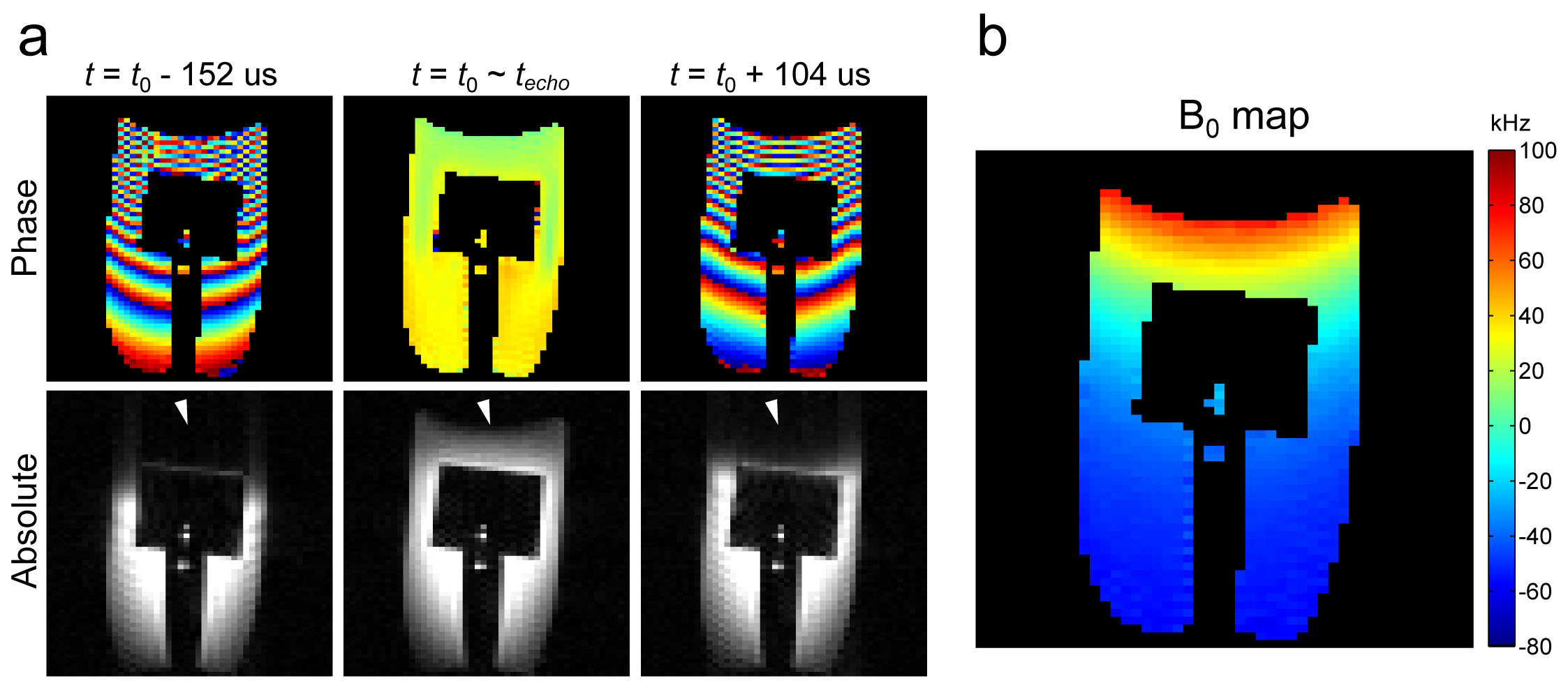
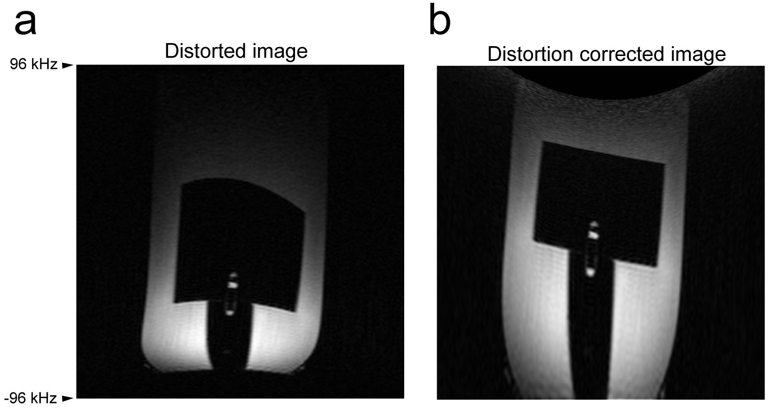
Phase images from the B0 mapping sequence clearly shows spin isochromats refocusing toward the echo peak and dephasing afterward; spin phase almost completely refocused at the echo peak (Fig.2a). Signals around the top of the phantom faded away during 100-200 μs from the refocusing echo peak due to intra-voxel dephasing (decoherence). The measured B0 map showed 180 kHz frequency variation over the phantom and a quadratically changing field along the z direction (Fig.2b). An image acquired with the nonlinear field gradient for spatial encoding resulted in severe image distortion in a reconstructed image; straight edges of Teflon object inside of the phantom are highly distorted (Fig.3a). The image distortion was nicely corrected according to the distortion field calculated from the measured B0 map (Fig.3b) as compared to the low resolution distortion-free images in Fig.2. While there is stronger signal intensity gradient along the vertical direction in the distorted image, the intensity gradient is mitigated with intensity correction using Jacobian in the corrected image.Discussion
The proposed method uses refocused echoes generated by the MP-SSFP sequence. A similar B0 mapping using chemical shift imaging (CSI) with free induction decay acquisition may be possible. However, under extremely inhomogeneous fields like the setting in this study, CSI-based B0 mapping is also highly challenging. CSI needs at least a few hundred microsecond delay between excitation and data acquisition for phase encoding gradient, which is long enough for signals in steep gradient regions to fade away as shown in Fig.2.
In this study, we demonstrated image distortion and intensity correction using a measured B0 map. However, there is still some residual signal intensity drop toward the top of the phantom observed in the corrected image. The signal intensity drop is mainly from spatially changing diffusion weighting, since diffusion contrasts are dependent on local field gradients; there is steeper field gradient around the top of the phantom (i.e. stronger diffusion weighting).
Conclusions
The proposed B0 mapping using MP-SSFP can measure inhomogeneous B0 fields with an order of hundred kHz off-resonance.Acknowledgements
This study was supported by NIH grants R24MH105998, P41EB015894, S10RR023730 and S10RR027290.References
1. Patz, S., S.T. Wong, and M.S. Roos, Missing pulse steady-state free precession. Magn Reson Med, 1989. 10(2): p. 194-209.
2. Yoo, T.S., et al., Engineering and algorithm design for an image processing Api: a technical report on ITK--the Insight Toolkit. Stud Health Technol Inform, 2002. 85: p. 586-92.
Figures