3967
Comparison of R2* and B0 Field in 2D and 3D Ferumoxytol-Enhanced Chemical Shift-Encoded MRI of the Healthy Rhesus Placenta1Biomedical Engineering, University of Wisconsin-Madison, Madison, WI, United States, 2Radiology, University of Wisconsin-Madison, Madison, WI, United States, 3Global MR Applications and Workflow, GE Healthcare, Menlo Park, CA, United States, 4Comparative Biosciences, University of Wisconsin-Madison, Madison, WI, United States, 5Medical Physics, University of Wisconsin-Madison, Madison, WI, United States, 6Obstetrics and Gynecology, University of Wisconsin-Madison, Madison, WI, United States, 7Wisconsin National Primate Research Center, University of Wisconsin-Madison, Madison, WI, United States, 8Medicine, University of Wisconsin-Madison, Madison, WI, United States, 9Emergency Medicine, University of Wisconsin-Madison, Madison, WI, United States
Synopsis
A motion-robust 2D sequential chemical shift-encoded MRI (CSE-MRI) technique with a short temporal footprint for each slice is investigated for ferumoxytol-enhanced MRI of placental inflammation. In this study, the proposed 2D technique was compared with the reference 3D-CSE-MRI technique in healthy pregnant rhesus, which were anesthetized eliminating fetal motion. B0 field map boundary measurements, as well as R2* measurements in the placenta were compared across the two techniques to assess the accuracy of the 2D technique. High correlations between the measurements from 2D-CSE-MRI and 3D-CSE-MRI were demonstrated and thus provide promise for motion-robust imaging of human placental inflammation.
Introduction
In the pathologies of preeclampsia, the altered immune cell (e.g. macrophage) activation and distribution at implantation may play a critical role1. The FDA-approved iron supplement ferumoxytol is an ultra-small superparamagnetic iron oxide that is phagocytosed by macrophages. Therefore, ferumoxytol-enhanced MRI in the placenta may enable safe and sensitive detection of increased macrophage density in inflammation2. Previously proposed 3D chemical shift-encoded (CSE) MRI enables R2*- and B0- mapping, both of which may enable assessment of tissue ferumoxytol concentration3. However, maternal respiration and fetal motion in the uterus may lead to artifacts in 3D imaging due to the long temporal footprint (~5 minutes), precluding the accurate estimation of R2* and B0 field maps.
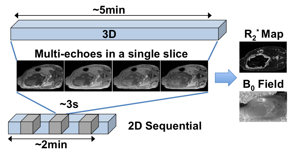
The proposed 2D sequential CSE-MRI has a small temporal footprint (e.g. ~3 seconds for each slice as shown in Figure 1), which may enable robust imaging even in the presence of fetal motion. However, it is unknown whether the 2D sequential technique enables accurate R2* and B0 field mapping. In this study, we compared the estimated R2* and B0 field in 2D-CSE-MRI to the standard 3D-CSE-MRI in anesthetized healthy pregnant rhesus. In this animal model, fetal motion is suppressed due to the anesthesia, therefore enabling reliable imaging with the standard 3D-CSE-MRI technique. For this reason, this animal model provides a unique opportunity to validate the proposed 2D-CSE-MRI technique, which may be the only reliable alternative for imaging in human placenta in the presence of fetal motion.
Methods
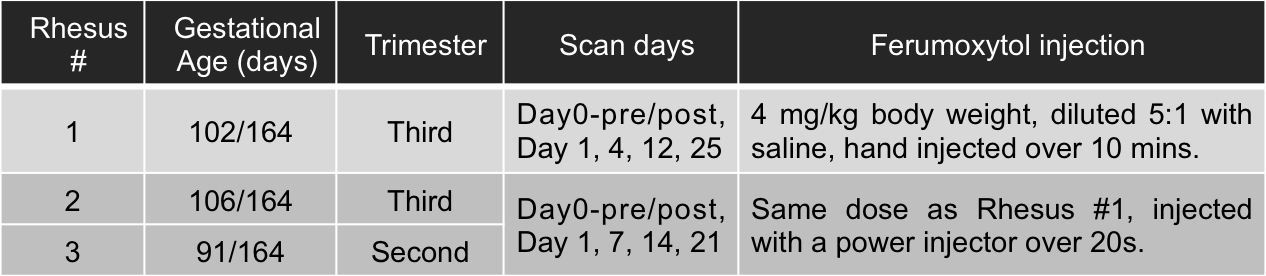
All procedures were approved by our institution’s animal care and use committee (IACUC). Three healthy pregnant rhesus monkeys were imaged on a clinical 3.0T MRI system (GE Healthcare Discovery MR 750, Waukesha, WI) using a 32-channel phased array torso coil (Neocoil, Pewaukee, WI). All mother rhesus monkeys were under general anesthesia during the scanning, which also anesthetized the fetus, thereby eliminating fetal motion. Six time points were collected for each monkey. Details on the gestational age of the animals, the scan days and ferumoxytol injection are shown in Table 1.
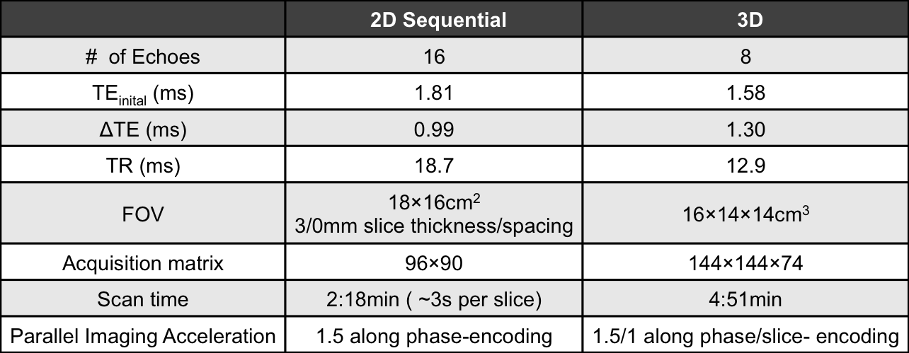
A multi-echo spoiled gradient-echo 2D sequential acquisition without respiratory gating was optimized for motion robustness and performed in the placenta of each animal. A multi-echo spoiled gradient-echo 3D acquisition with respiratory gating was acquired as the reference for ferumoxytol-enhanced CSE MRI in the placenta. Scan parameters are listed in Table 2. R2* and B0 field maps were obtained by using a CSE reconstruction4. R2* measurements were performed using ROIs drawn in two separate placental lobes of each animal at each time point. B0 field values in the placenta and adjacent amniotic fluid (AF) of each monkey (at day 0-post and day 1) were measured and subtracted to obtain the boundary B0 field difference (ΔB0, obtained along boundaries parallel to the main magnetic field), a measure of magnetic susceptibility difference between the two regions3. Linear regression analysis was used to assess the correlation of R2* as well as ΔB0 estimated in 2D versus 3D imaging.
Results
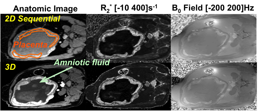
Anatomic images, and R2* and B0 field maps from 2D- and 3D-CSE-MRI of the third rhesus at day 0-post are shown in Figure 2. No obvious motion artifacts are shown in 2D sequential imaging, which demonstrates the motion robustness of this technique. Figures. 3a and 3b show highly consistent R2* measurements in 2D and 3D imaging with a slope of 0.92 (R2=0.979) in the linear regression on R2* and a slope of 0.89 (R2=0.988) in the linear regression on log(R2*). The boundary B0 field differences ΔB0 of all three rhesus monkeys at day 0-post and day 1, and the corresponding linear regression are shown in Figure 3c. The slope of the linear fitted line is close to 1 (1.13 with R2=0.989), indicating that the susceptibility difference between placenta and adjacent amniotic fluid could be consistently estimated from ΔB0 in both 2D- and 3D-CSE-MRI.Discussions and Conclusions
In this study, we have demonstrated the accuracy of 2D-CSE-MRI for quantitative ferumoxytol-enhanced MRI in the healthy rhesus placenta. Excellent image quality demonstrated motion robustness of 2D-CSE-MRI without the need for respiratory gating due to the short temporal footprint in imaging. 2D acquisitions demonstrated good agreement relative to the standard 3D acquisitions, which demonstrated good accuracy of R2* and B0 field mapping in 2D imaging. Upon further validation in inflamed rhesus model and in human scans, 2D ferumoxytol-enhanced CSE-MRI may provide a reliable approach for the evaluation of inflammation in human placenta in the presence of fetal motion.Acknowledgements
The authors acknowledge the support of the NIH (grants U01-HD087216, R01-DK100651, K24-DK102595, and P51-OD011106). We also thank GE Healthcare for their support.References
1. Faas MM, et al. Monocytes and macrophages in pregnancy and pre-eclampsia. Front Immunol. 2014; 5:298.
2. Neuwelt A, et al. Iron-based superparamagnetic nanoparticle contrast agents for MRI of infection and inflammation. AJR. 2015; 204(3):W302.
3. Hernando D, et al. Magnetic susceptibility as a B0 field strength independent MRI biomarker of liver iron overload. Magn Reson Med. 2013; 70(3):648-656.
4. Yu H, et al. Multiecho water-fat separation and simultaneous R2* estimation with multifrequency fat spectrum modeling. Magn Reson Med. 2008; 60(5):1122-1134.
Figures