3966
Reference-Free Distortion Correction with Segmented Partial Fourier Acquisitions for High Resolution DTI at 7T1Dept. of Radiology, Medical Physics, University Medical Center Freiburg, Freiburg, Germany, 2Department of Cognitive Neuroscience, Maastricht Brain Imaging Centre (MBIC)
Synopsis
Single-Shot diffusion weighted echo planar imaging (EPI) is known for its strong distortions due to long imaging readouts. However, even for segmented acquisitions, high resolution diffusion weighted imaging suffers from image distortions. Our approach shows that intrinsic field information of a segmented DTI acquisition can be used for robust distortion correction without blurring effects. In addition this approach offers the advantage to reduce artifacts from partial Fourier (PF) acquisitions due to better data distribution in k-space. In combination with the MUSE approach, this promising technique is applied to whole brain DTI with a resolution of 1mm isotropic.
Introduction
Until today diffusion tensor imaging (DTI) in clinical research is mostly restricted to single-shot EPI, which substantially limits the achievable resolution and suffers from strong image distortions. Recently, navigated segmented acquisitions (e.g. Resolve) became more popular and allow for much higher resolution. In contrast to the navigated approach, we used multiplexed sensitivity encoding (MUSE, [1]) to overcome phase-inconsistencies between segments. In this work we show that image distortions can be further reduced, by inverting phase encode (PE) directions in different segments. These segments are then used to calculate a fieldmap of the acquired data without additional time penalty during acquisition. In combination with the MUSE approach, this promising technique is applied to whole brain DTI with a resolution of 1mm isotropic.Methods
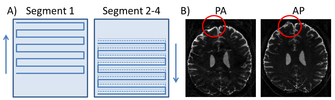
Sequence design: DWI-data was acquired on a 7T Scanner (Siemens Healthcare) with a Stejskal-Tanner single spin echo sequence (TR/TE: 14600/59ms, diffusion weighting: 1000s/mm2, 30 directions, matrix: 192x192x115, 1mm isotropic, PF 6/8). Segmentation was performed in PE-direction using 4 interleaved segments. The protocol was run twice, once with the PE direction in Anterior-Posterior (AP) in each segment and once with the PE direction flipped (PA) in the first segment relative to the other three segments (AP) as displayed in figure 1A. In addition to the inverted PE direction, this has the effect, that for acquisitions with partial Fourier (PF), information on both sides of the k-space is acquired.
Muse reconstruction: Coil-sensitivities are calculated from the unweighted (b=0s/mm2) and unflipped data. These profiles are then used to reconstruct each under-sampled segment to determine the phase distribution after diffusion weighting. This phase is than incorporated in the sensitivity maps and a final reconstruction is performed using data from all four segments [1].
DicoFlip reconstruction: In addition, the images reconstructed from two segments of the second dataset (acquired AP and PA, figure 1B) are used to calculate a field-map [2]. This is then included in the final reconstruction defining the full forward-operator as described in [3]:
$$$ S_{(j,l)} (k_x,k_y )=∫ c_{(j,l)} (r) e^{(-sg(l)iω(r) k_x-i(sg(l) k_x r_x+ k_y r_y))} ρ(r)dr^3 $$$
$$$S_{(j,l)}$$$ is the signal coming from the j-th coil and l-th segment. $$$sg(l)$$$ denotes the sign indicating bottom-up/top-down traversal. The sensitivities $$$c_{(j,l)} (r)$$$ are the modified coil-sensitivities (incorporating the phase distribution) calculated from the j-th coil and l-th segment.
Results
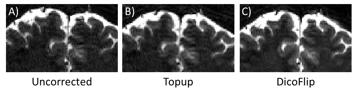
Figure 2 shows one exemplary slice without diffusion weighting (b=0s/mm2).
a) First dataset with MUSE reconstruction, without distortion correction
b) Second dataset with distortion correction (Topup)
c) Second dataset with DicoFlip reconstruction
Both correction methods reduce distortions as displayed in the enlarged image-segment. However, the Topup reconstruction suffers from slight blurring, which can be overcome with the new DicoFlip reconstruction.
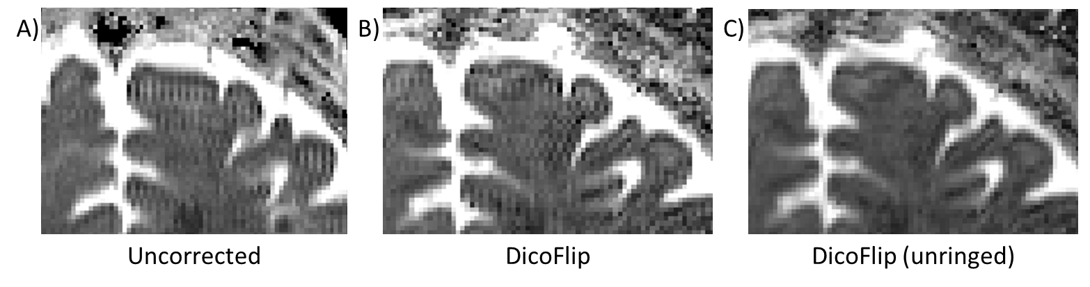
Figure 3 displays the DTI data (mean diffusivity) calculated from all data reconstructed without (A) and with DicoFlip (B). A) The unflipped data shows ringing in only one dimension (readout), the ringing artifact in the PE direction is blurred out. B) The data acquired with DicoFlip maintains this ringing in both dimensions. However, in contrast to the PF artifact, this ringing can be corrected using an additional post-processing step [4]. The result after unringing is shown in C).
Discussion and Conclusion
Our approach shows that intrinsic field information of a
segmented DTI acquisition can be used for robust distortion correction without
blurring effects. In addition to the time savings for additional reference
scans, this stands in clear contrast to established methods where the
distortion correction is performed in additional post-processing steps. For
acquisitions with PF, it was shown that the improved data distribution in
k-space leads to a substantial reduction of PF-artifacts. Combined with MUSE, DicoFlip
allowed for distortion free high resolution DTI at ultra-high field.Acknowledgements
References
[1] N. Chen, A. Guidon, H.-C. Chang, und A. W. Song, „A robust multi-shot scan strategy for high-resolution diffusion weighted MRI enabled by multiplexed sensitivity-encoding (MUSE)“, NeuroImage, Bd. 72, S. 41–47, Mai 2013.
[2] J. L. R. Andersson, S. Skare, und J. Ashburner, „How to correct susceptibility distortions in spin-echo echo-planar images: application to diffusion tensor imaging“, NeuroImage, Bd. 20, Nr. 2, S. 870–888, Okt. 2003.
[3] M. Reisert and M. Herbst, „Reference-free Distortion Correction for EPI by Flipped k-space Segments (DICOFLIP)“. ISMRM Proceedings, 2015.
[4] E. Kellner, B. Dhital, V. G. Kiselev, und M. Reisert, „Gibbs-ringing artifact removal based on local subvoxel-shifts: Gibbs-Ringing Artifact Removal“, Magn. Reson. Med., Bd. 76, Nr. 5, S. 1574–1581, Nov. 2016.
Figures