3963
Partial Fourier techniques in single-shot cross-term spatiotemporal encoding (xSPEN) MRI1Weizmann Institute of Science, Rehovot, Israel
Synopsis
xSPEN is a new single-shot imaging approach with exceptional resilience to field heterogeneities: its images do not suffer from miss-registrations, require a priori information nor use post-acquisition corrections, to deliver faithfully the spins’ spatial distribution. xSPEN, however, suffers from SNR penalties due to its non-Fourier nature and its considerable diffusion losses –especially when desiring high resolution. This study introduces partial Fourier transform approaches that acting along either the readout or the spatiotemporally-encoded dimensions, reduce both of these penalties. The principles of these partial FT methods are given, and applications in materials, preclinical and human single-shot xSPEN imaging are presented.
Purpose
To reduce the strong diffusion losses and shorten the minimum TE of single-shot xSPEN,1 thereby improving trade-offs between resolution and SNRIntroduction
Central to fast MRI is EPI,2 a single-shot approach that rapidly scans k-space. Recently we introduced cross-term spatiotemporal encoding –xSPEN for short; a single-shot approach which presents a remarkable resilience to field inhomogeneities and/or multiple chemical sites.1 By forfeiting the FT, however, xSPEN presents SNR penalties vis-à-vis EPI or SPEN3–especially when seeking high resolution. This study introduces partial FT (pFT) techniques that acting along either the readout or SPEN dimensions, improve xSPEN’s resolution and SNR.Methods
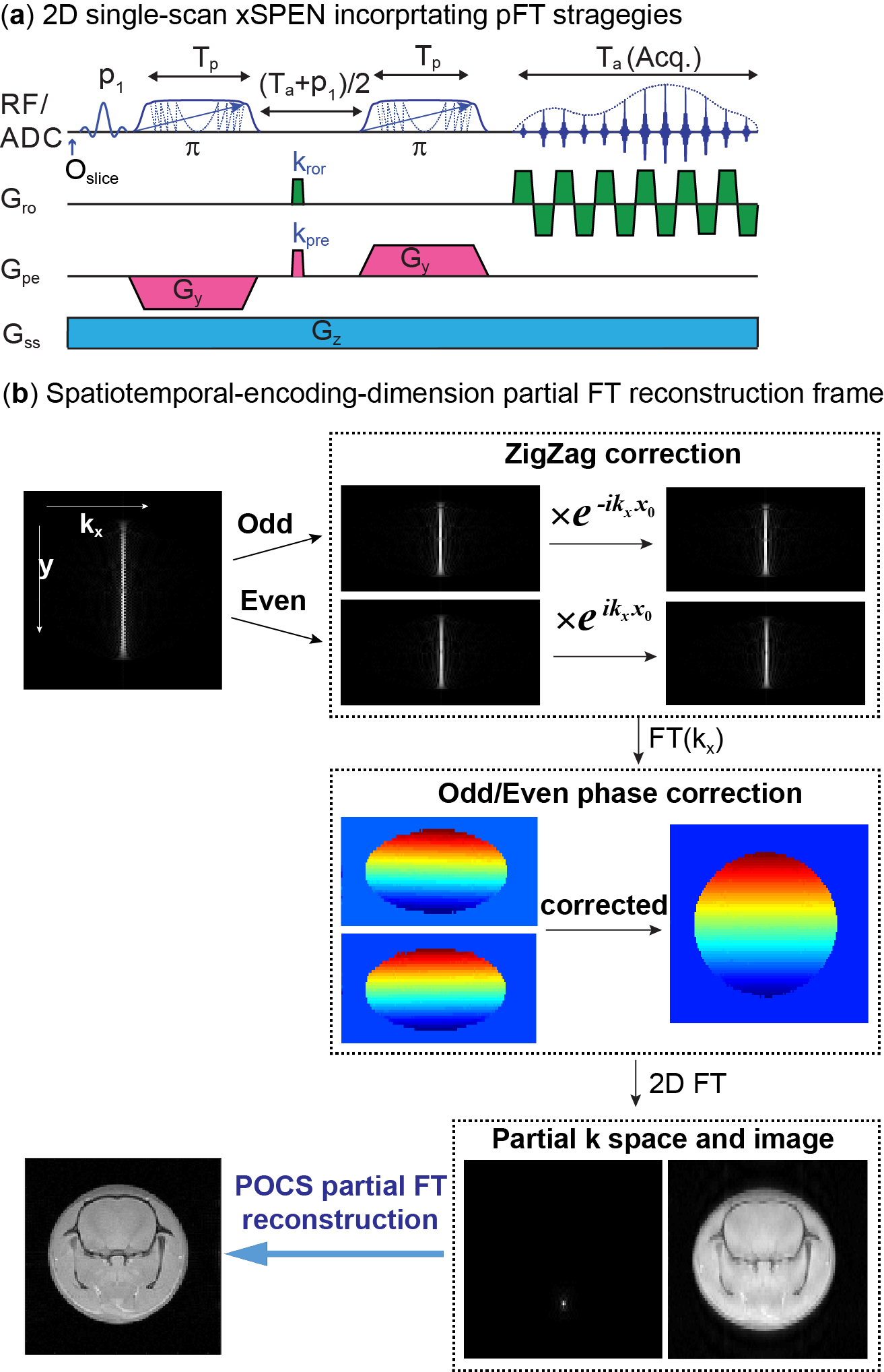
xSPEN applies frequency-swept pulses under the action of bipolar y- and constant z-gradients imparting a preacquisition hyperbolic phase eiCyz (Fig. 1a), and applies during acquisition a constant z-gradient performing an “analog FT” on these data, and directly delivering a y-axis image. Since these y-axis images are read out under continuously spin-echoed, inhomogeneity-free conditions, their inverse FT is the equivalent of an ideal, ky-encoded FID. It is known that the inherent resolution of such k-space data depends on kymax; partial FT procedures use this to enhance resolution without extending acquisition times, by shifting the k-echo and placing it asymmetrically within the acquisition window4. To enjoy a similar advantage in xSPEN we introduce a short prephasing pulsed gradient along y (kpre in Fig. 1a) that phase modulates the acquired y image. In practice, the zigzag effect associated with the oscillating Gx and constant Gz gradients acting during xSPEN’s 2D single-shot acquisition, needs to be taken care of before applying a pFTy. The reconstruction procedure that we developed for this is summarized in Fig. 1b. In addition, a prephasing along the x-axis (kror in Fig. 1a) yields the option of applying a pFTx. Such reconstruction can be separately done on positive and negative kx-axis acquisitions, and the two datasets combined in image space without suffering from phase problems. As both pFTx and PFTy can shorten the TE and overall duration of the Gz-gradient application, they both help reduce diffusion and T2 losses.Results and Discussion
Phantom and in-vivo mice scans were carried out using an Agilent 7T scanner; volunteer scans were carried out in a Siemens 3T.
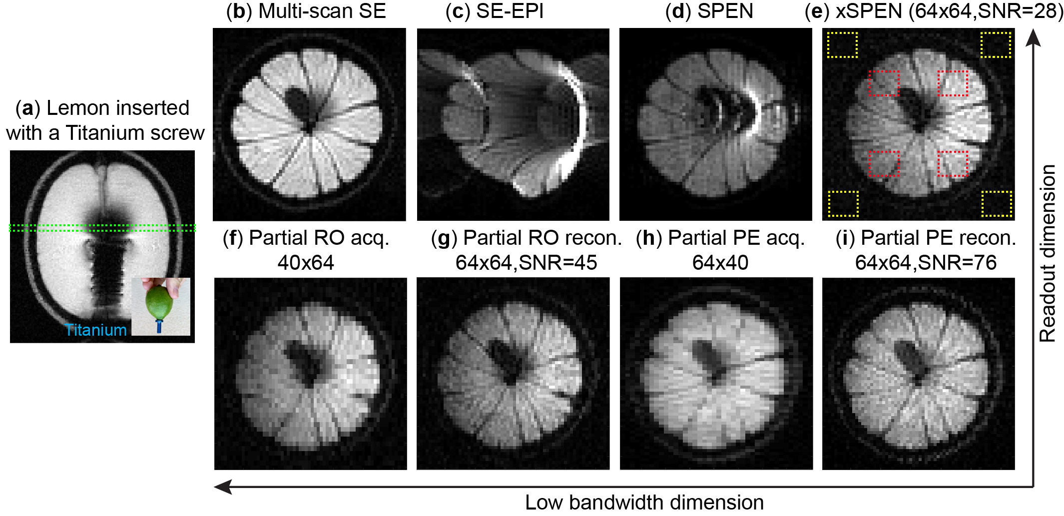
Figure 2 shows results from a titanium screw inserted inside a lemon. Single-shot xSPEN without (Fig. 2e) or with pFT (Fig. 2f-i) yields considerably more truthful images than EPI and SPEN acquisitions. While both pFTx (option I, Fig. 2g) and pFTy (option II, Fig. 2i) give better SNRs than the original single-shot xSPEN (Fig. 2e) under same resolutions, the phase-encoded option (Fig. 2i) results in better SNR due to its larger reduction in the overall Gz duration.
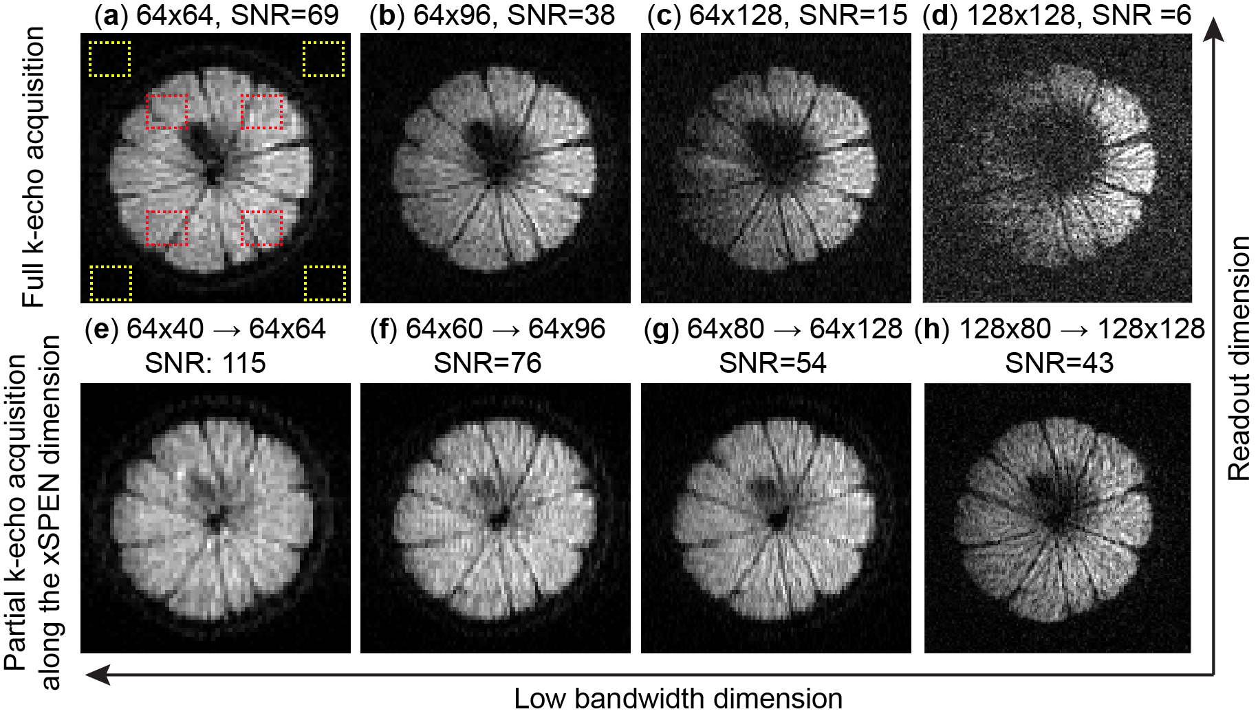
Figure 3 demonstrates how xSPEN’s pFT improves the trade-off between resolution and SNR. SNR is increasingly degraded when higher resolution images are acquired, due to xSPEN’s enhanced diffusion losses (Fig. 3a-d). Images reconstructed using pFTy (Fig. 3e-h) not only show better SNRs than their conventional xSPEN counterparts; the higher the resolution desired, the larger the SNR benefit arising from pFT. This can be understood due to the latter’s attenuation of the diffusion-derived losses, which increase in proportion to an exponential power of the gradient’s duration.
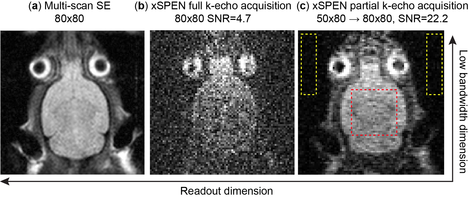
Figure 4 shows mice results illustrating pFT’s advantages in single-shot in vivo xSPEN imaging. Notice the SNR improvements brought about by the pFT (>5x) for the 300 µm in-plane resolution targeted, as well as the absence of distortions (e.g., near eyeballs and ears).
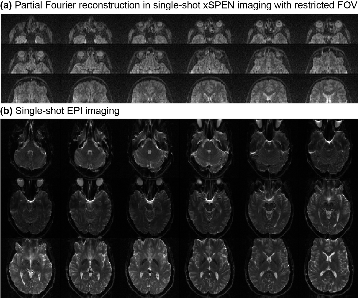
Figure 5 illustrates a similar advantage, but for a human frontal orbital region. Due to the sinuses and eye sockets, single-shot EPI suffers here from serve distortions (Fig. 5b). xSPEN yields distortion free images for this region, but strong diffusion losses when in-plane resolution is better than 2×2 mm2 render this approach impractical. On the other hand, Figure 5a shows how single-shot xSPEN images using pFTy, can successfully tackle these regions with a restricted FOV.
Conclusions
pFT single-shot xSPEN improves SNR, delivering higher spatial resolutions while preserving the method’s unprecedented resilience to field inhomogeneities. This can help to expand the potential applications of this emerging single-shot imaging technique.Acknowledgements
Funding by grants ISF #795/13, ERC-2014-PoC # 633888, Minerva Foundation #712277, the Kimmel Institute of Magnetic Resonance (Weizmann Institute). ZZ thanks Israel’s Council of Higher Education and Koshland Foundation for fellowships. We are also grateful to Dr. Sagit Shushan (Wolfson Medical Center), and the Weizmann MRI team (Edna Furman-Haran, Fanny Attar and Nachum Stern).References
1 Zhang Z., Seginer A., Frydman L., Single scan MRI with exceptional resilience to field heterogeneities. Magn Reson Med; doi:10.1002/mrm.26145. 2 Mansfield P., Multi-planar image-formation using NMR spin echoes. J Phys C: Solid State Phys 1977;10:L55-L58. 3 Schmidt R., Frydman L., New spatiotemporal approaches for fully refocused, multislice ultrafast 2D MRI. Magn Reson Med 2014;71:711-722. 4 Liang Z.P., Lauterbur P.C., Principles of magnetic resonance imaging: a signal processing approach, IEEE Press, 2000. 5 Haacke, E. M., Lindskogj E. D. and Lin W., A fast, iterative, partial-Fourier technique capable of local phase recovery. J Magn Reson 1991;92:126-145.
Figures