3939
Image-Based Non-Rigid Motion Correction for Free-breathing 3D Abdominal MRI1Academy for Advanced Interdisciplinary Studies, Peking University, Beijing, People's Republic of China, 2Philips Healthcare, Suzhou, People's Republic of China, 3College of Engineering, Peking University, Beijing, People's Republic of China, 4Department of Radiology, Peking University First Hospital, Beijing, People's Republic of China
Synopsis
Free breathing abdomen imaging requires non-rigid motion registration of the unavoidable respiratory motion in the 3D under-sampled datasets. In this work, pyramidal Lucas-Kanade based optical flow estimation is proposed to perform upper abdomen registration, which enables reconstruction of motion-free abdominal images throughout the respiratory cycle. Preliminary results on images acquired using 3D golden radial phase encoding eThrive scan demonstrate that our approach makes vessels in liver sharper and more consecutive when compared to traditional NMC and LREG based methods.
Introduction
Upper abdomen MRI is affected by respiratory motion which are commonly compensated by breath-holding[1]. However, free-breathing acquisition is preferred and even required in patients with limited breath hold capability.
Golden Angle radial [2] sampling has favorable properties for free-breathing MRI, including motion robustness and flexible reconstruction options.
In this work, we propose the combination of a self-gated 3D Golden Angle radial data acquisition scheme with pyramidal Lucas-Kanade based optical flow estimation to obtain motion-free abdominal images throughout the respiratory cycle.
Method
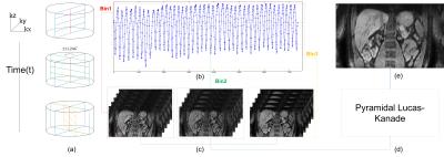
1. Data Acquisition MRI scans of 9 healthy volunteers were perfomed on a clininal 1.5T MRI system (Ingenia, Philips Healthcare, Best, The Netherlands). MR data acquisition is performed using a 3D golden radial phase encoding eThrive sequence, where Cartesian frequency encoding is used along the partition dimension (kz) and golden-angle (θ=111.246º) radial phase encoding is used in the kx -ky plane (Figure 1a). Relevant imaging parameters of included: slice thickness = 3 mm with over contiguous sampling ; flip angle = 10 degrees; field of view (FOV) = 450 × 450 × 249 mm2; sense factor along z = 1.41; number of readout points in each spoke = 752 with two times oversampling; spatial resolution = 1.00 ×1.00 ×3.00 mm3; repetition time/echo time (TR/TE) = 4.88 ms/2.06 ms. A total of 751 spokes were acquired for each partition, with a total scan time of ~180 s.
2. Data Binning In our work, an adaptive method was used to estimate respiratory signal. The 1D fast Fourier transform along the feet-head (FH) direction to the center k-space profiles was applied to compute the projection profiles of the 3D volume. The respiratory motion detection was performed by first aligning projection profiles into a 2D image (Fig 1b), followed by envelope extraction of the image. Based on the respiratory motion signal, the continuously acquired golden-angle radial datasets were divided into three respiratory bins (Fig 1c), in which spokes were in the same motion position, similar to [3].
3. Reconstruction of Respiratory Bins Reconstruction was developed and performed using Recon2.0 platform by Philips. A 3D under-sampled image for each bin was reconstructed by gridding[4] and followed by SENSE to unfold the warped image along F-H direction. It took ~80 seconds to reconstruct a 3D volume using workstation equipped with 16GB DDR RAM and 2 intel XEON E5-1620 CPUs.
4. Motion Modeling Unlike Buerger et al[5], where LREG tool was used to register reconstructed bin images to a common respiratory position, here we propose to apply pyramidal lucas-kanade registration algorithm (Fig 1d).
The proposed method was compared with the non-compensated approach and reconstruction framework of in terms of signal-to-noise ratio (SNR), image sharpness, visual image scoring and registration time.
Result
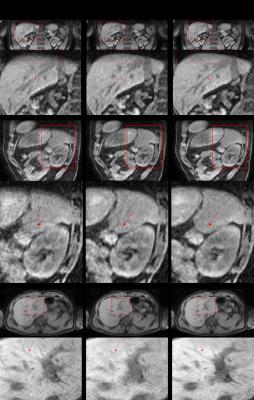
Multiple slice orientations for the non-motion corrected (NMC), LREG based reconstruction and PLK based reconstruction for volunteer 1 is shown in figure 3. The coronal images indicate that LREG has caused fracture in vessel, while vessels of PLK are sharper and more consecutive. As shown in sagittal view, LREG has distorted and blurred the liver contours compared with NMC and LREG. The zoom-in axial images present that PLK would lead to vessel deformed in noisy region. However, PLK could make vessel delineated more clearly.
Statistical results of the signal-noise-ratio, visual score, vessel sharpness and registration time over all volunteers are shown in Table 1. The SNR indicates that the PLKR have the best image quality (267.42±96.73), significantly better than NMC (116.67±44.70) and LREG (187.93±96.68). Image visual score agreed with SNR, marking PLK (3.85±0.12) the best, followed by LREG (3.43±0.13) and NMC (2.55±0.09). Vessel sharpness assessment yielded similar values between the PLK (0.81±0.03) and LREG (0.80±0.04), differentiating them from the NMC (0.78±0.06). When comparing with the LREG based reconstruction, the PLK based reconstruction reduces the registration time from 1h to 0.5h.
Discussion and Conclusion
We have presented a novel technique based on pyramidal Lucas-Kanade, which enables reconstruction of motion-free abdominal images throughout the respiratory cycle. Our preliminary result demonstrated that our scheme are superior to NMC and LREG based methods. The described technique offers several potential advantages: high image quality with both high SNR, visual score and vessel sharpness; time-efficient registration. The presented technique could also be extended to simplify and improve dynamic contrast-enhanced abdomen exams.Acknowledgements
No acknowledgement found.References
1. Bernstein, M.A., K.F. King, and X.J. Zhou, Handbook of MRI pulse sequences. 2004: Elsevier.
2. Stehning, C., et al., Free- breathing whole- heart coronary MRA with 3D radial SSFP and self-navigated image reconstruction. Magnetic resonance in medicine, 2005. 54(2): p. 476-480.
3. Buerger, C. and C. Prieto, Highly efficient 3D motion-compensated abdomen MRI from undersampled golden-RPE acquisitions. Magma Magnetic Resonance Materials in Physics Biology & Medicine, 2013. 26(5): p. 419-29.
4. Beatty, P.J., D.G. Nishimura, and J.M. Pauly, Rapid gridding reconstruction with a minimal oversampling ratio. IEEE transactions on medical imaging, 2005. 24(6): p. 799-808.
5. Buerger, C., et al., Nonrigid motion modeling of the liver from 3-D undersampled self-gated golden-radial phase encoded MRI. IEEE Transactions on Medical Imaging, 2012. 31(3): p. 805-15.
Figures


