3935
One-Dimensional Phase Navigation of Diffusion-Weighted 3D TSE for High Resolution Musculoskeletal Diffusion Imaging1Department of Diagnostic and Inteventional Radiology, Technical University of Munich, Munich, Germany, 2IMETUM, Technical University of Munich, Garching, Germany, 3Philips Healthcare, Hamburg, Germany, 4Philips Healthcare, Best, Netherlands, 5Department of Neuroradiology, Technical University of Munich, Munich, Germany
Synopsis
Diffusion-prepared 3D TSE (dprep-3D-TSE) imaging has been recently applied for high-resolution distortion-free body and musculoskeletal DWI. Dprep-3D-TSE has been combined with magnitude stabilizers to reduce magnitude modulation effects induced by both motion and eddy current effects. In addition, velocity compensation has been proposed to reduce motion-induced phase modulation effects. However, given that dprep-3D-TSE is a multi-shot diffusion technique, it suffers from motion-induced phase variation effects across different shots and it requires phase navigation. The purpose of the present study is to develop an acquisition and phase correction scheme for one-dimensional phase navigation of dprep-3D-TSE imaging.
Purpose
Diffusion-prepared TSE (dprep-TSE) sequences have been recently emerging for body and musculoskeletal diffusion-weighted (DW) imaging, due to their ability to achieve higher spatial resolution without being sensitive to the geometric distortions of the typically used single-shot echo planar imaging1-4. Dprep-TSE sequences have been also combined with 3D imaging to achieve isotropic resolution DW images. However, phase errors induced by motion and eddy current effects during the diffusion encoding period affect not only the magnitude but also the phase of the dprep-3D-TSE signal5. The use of magnitude stabilizers removes the magnitude modulation induced by eddy currents and motion6 and velocity compensation reduces motion-induced phase modulation. However, the variation of the remaining motion-induced phase modulation across different k-space segments (shots) remains a significant source of artifacts and diffusion parameters quantification errors. The purpose of the present work is to develop a phase-navigated DW 3D TSE sequence, using a one-dimensional phase navigator acquired during the start-up echoes of the 3D TSE flip angle train and to show the reduction of the associated phase errors in two musculoskeletal tissues.Methods
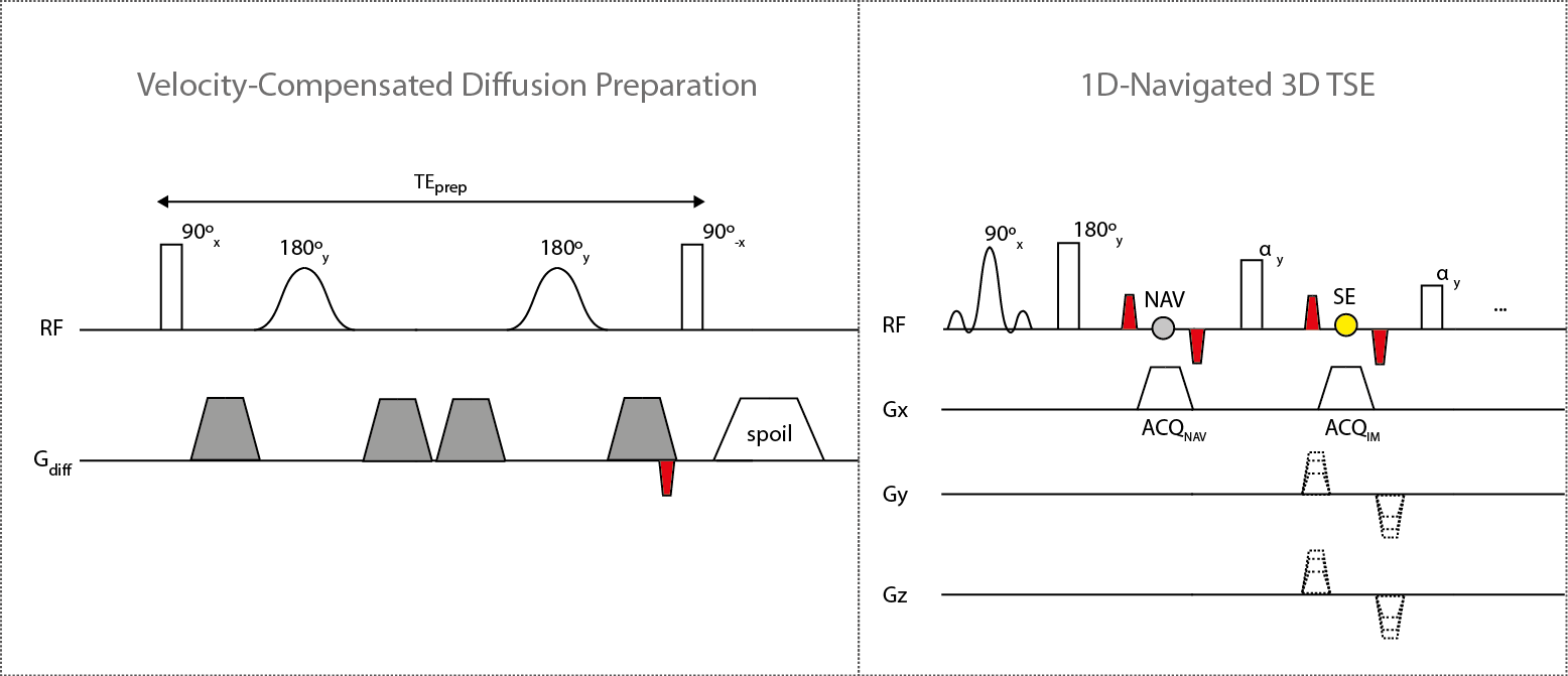
Sequence development: The used diffusion preparation consisted of an RF component of double-refocused hyperbolic secant pulses, for minimizing sensitivity to transmit B1 effects, and of a velocity-compensated diffusion gradient configuration for minimizing motion effects6. A single dephasing gradient (magnitude stabilizer) was placed before signal restoration and later balanced in the 3D TSE readout for minimizing phase errors induced by motion and eddy currents. 3D TSE readout consisted of the acquisition of imaging echoes using phase encoding in ky and kz directions while a navigator echo was acquired for each individual shot with no ky or kz phase encoding.
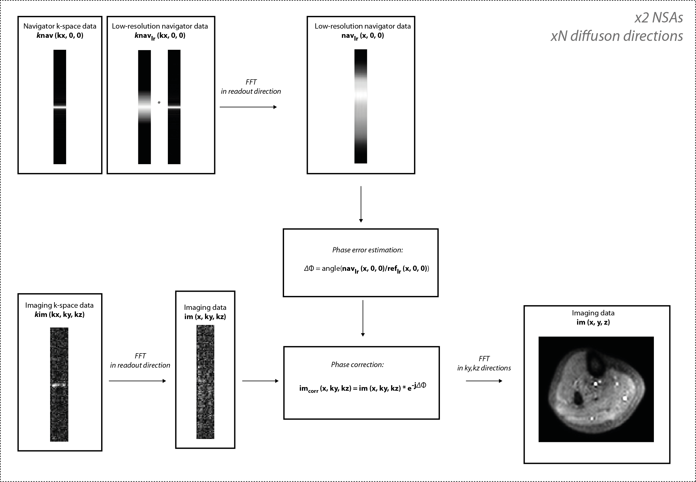
1D phase correction and reconstruction: Low-resolution k-space navigator data knavlr(kx,0,0) was Fourier-transformed in the readout direction and the phase of the low-resolution image-space navigator navlr(x,0,0) of each diffusion direction was compared to the phase of the low-resolution image-space b0 navigator reflr(x,0,0). The estimated phase difference ΔΦ was used to phase-correct each corresponding diffusion direction of hybrid-space imaging data im(x,ky,kz) by means of complex multiplication. The corrected imaging data imcorr(x,ky,kz) was 2D Fourier-transformed in ky and kz directions and carried through regular reconstruction. The above procedure was performed separately for individual NSAs and the two NSAs were averaged for removing FID artifacts.
In vivo measurements: DTI of the calf muscle of three healthy volunteers was conducted using a 16-channel knee coil and DTI of the lumbar plexus of two healthy volunteers was performed using a 16-channel torso coil and the integrated posterior coil. Both measurements were performed on a 3T Philips system with the developed sequence. Sequence parameters for muscle DTI: FOV=140×156×51mm3, acquisition voxel=2×2×6mm3, TR/TE=2500/26ms, TSEfactor=53, averages=2, b-values=0,400, DTI directions=10, TEprep=53ms, duration=13m50s. Sequence parameters for plexus DTI: FOV=380×380×70mm3 acquisition voxel=2.5×2.5×2.5mm3, TR/TE=1800/29ms, TSEfactor=50, averages=2, b-values=0,400, DTI directions=6, duration=16m53s.
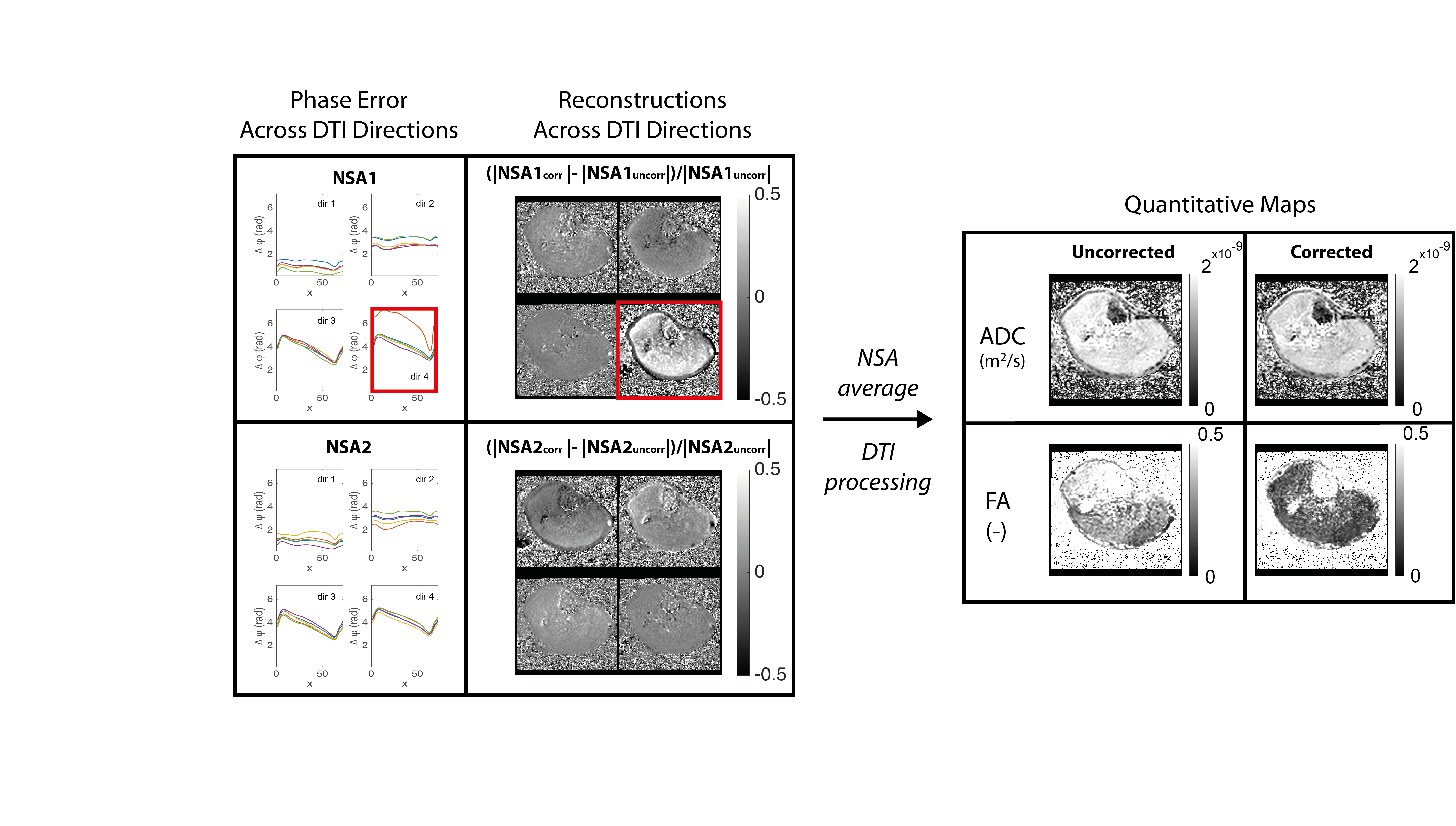
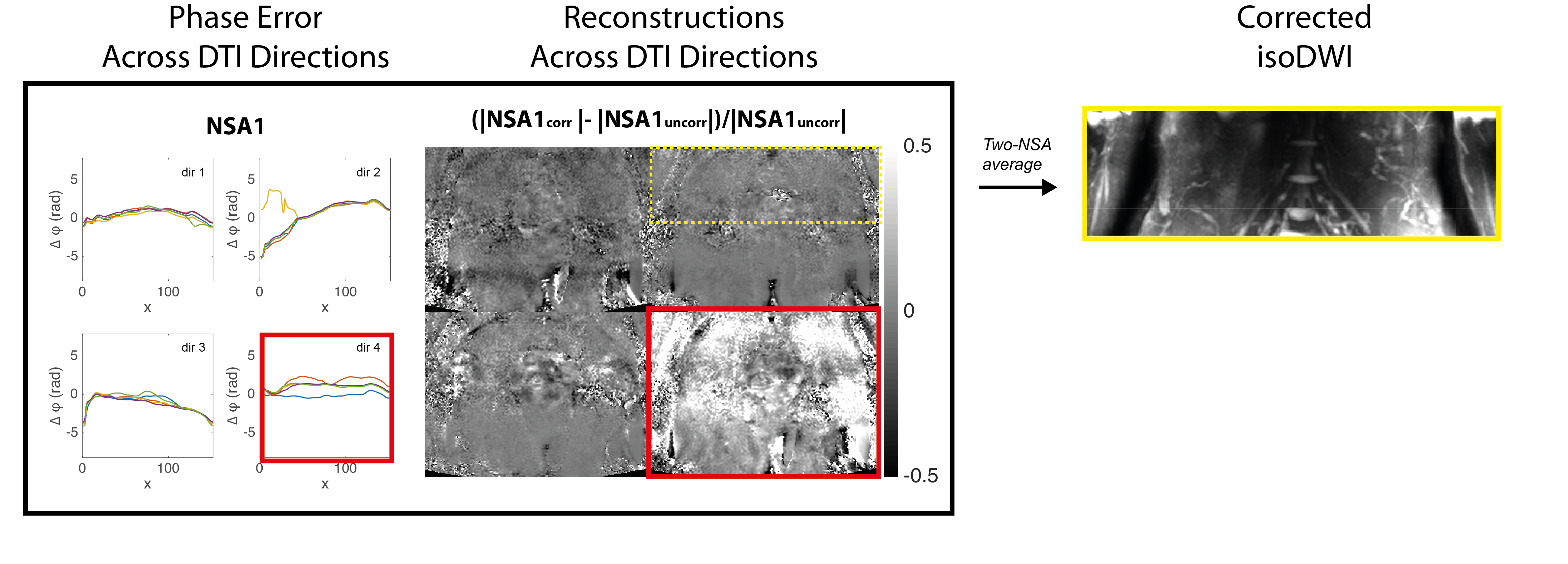
Post-processing: Mean ADC and FA maps were computed for muscle DTI data and iso-diffusion-weighted images were generated from the lumbar plexus data.
Results
Phase errors present in individual diffusion directions in muscle and lumbar plexus data (Figs.3,4) resulted in signal loss of the reconstructed images and thus in an overestimation of mean ADC and FA values for muscle data (Fig.3). Mean ADC and FA values measured for three subjects in the medial gastrocnemius muscle: ADCuncorr=(1.8±0.1)×10-9m2/s, ADCcorr=(1.6±0.08)×10-9m2/s, FAuncorr=0.42±0.05, FAcorr=0.21±0.01.Discussion & Conclusion
The presented measurement of intra-shot phase errors in 3D TSE show that motion-induced phase dispersion in individual diffusion acquisitions can result in significant signal loss, which in turn can result in erroneous quantification. 1D phase navigation and correction of diffusion-weighted 3D TSE data has been shown to reduce signal loss from motion-induced phase errors and to yield more accurate quantitative maps in calf muscle. In vivo results of the lumbar plexus show the presence of motion-induced phase errors in individual diffusion directions and phase-corrected diffusion-weighted images with high resolution are demonstrated to yield high signal and good delineation in small nerve roots and branches in regions affected by motion. The present work therefore introduces a phase-correction method that corrects for intra-shot motion-induced phase errors in diffusion-prepared 3D TSE and allows more accurate quantitative imaging using high isotropic resolution.Acknowledgements
The present work was supported by Philips Healthcare.References
1. Cervantes B, Zhang Q, van de Ven K, et al. High-resolution DTI of distal peripheral nerves using flow-compensated diffusion-prepared 3D TSE. Proc Intl Soc Mag Reson Med 2016;24:4530.
2. Nguyen C, Fan Z, Xie Y, et al. In vivo diffusion-tensor MRI of the human heart on a 3 tesla clinical scanner: An optimized second order (M2) motion compensated diffusion-preparation approach. Magn Reson Med 2016;76(5):1354-1363.
3. Xie Y, Yu W, Fan Z, et al. High resolution 3D diffusion cardiovascular magnetic resonance of carotid vessel wall to detect lipid core without contrast media. J Cardiovasc Magn Reson 2014;16:67.
4. Zhang Q, Cervantes B, Karampinos DC, et al. High resolution 3D diffusion imaging of carotid vessel wall using stimulated echo based diffusion prepared turbo spin echo sequence. Proc Intl Soc Mag Reson Med 2016;24:959.
5. Van AT, Cervantes B, Kooijman H, et al. How to minimize motion-induced phase and magnitude modulation in diffusion-prepared sequences? Proc Intl Soc Mag Reson Med 2016;24:4530.
6. Alsop DC. Phase insensitive preparation of single-shot RARE: application to diffusion imaging in humans. Magn Reson Med 1997;38(4):527-533.
Figures