3933
Effect of head motion on B0 shimming based on magnetic field probes1Radiology, Leiden University Medical Center, Leiden, Netherlands
Synopsis
Magnetic field probes (FP) can be used to monitor B0-changes and this approach has shown great potential for real time correction of artifacts produced by such changes. When estimating B0 fluctuations in the brain it is, however, not known how head motion influences the FP-based B0-estimation. Head motion introduces a B0-change both inside and outside the head, this study assesses the impact on field probe estimated B0-distributions within the brain at 7 Tesla. FP based correction after head motion can actually lead to a higher error especially evident when using third order spherical harmonics. Improved performance is, however, identified when using first order spherical harmonics.
Purpose
Magnetic field probes (FP) can be used to monitor B0-changes and this approach has shown great potential for real time correction of such changes by means of dynamic shimming. When estimating B0 fluctuations in the brain it is, however, not known how head motion influences the FP-based B0-estimation. Motion introduces a B0-change both inside and outside the head. It is unclear whether FP based B0-estimation still provide in such situations a correct estimation of the actual B0-changes within the brain. This study assesses the impact of head motion on field probe estimated B0-distributions within the brain. Correction with FP estimated B0 maps can actually lead to an increase in the error in B0 estimation. This is especially evident for third order spherical harmonics, while improved performance is identified for first order correction.Methods
All scans were performed on a 7 Tesla MRI (Philips Achieva, Best, The Netherlands). The study was approved by the local IRB and 5 healthy subjects were included after giving informed consent. The correspondence between FP-based and MR-measured B0 maps of the head was evaluated before and after changes in head position. The impact of head motion of FP-derived field estimation was assessed both by means of simulations based on a rotating head model as well as experimentally in volunteers actively performing head motion.
B0 field simulations, of a head model were used to estimate B0 maps both inside the head as well as surrounding the head to assess B0-changes at virtual FP positions. The simulated FP-signals were subsequently fitted to yield an estimation of the B0-distribution within the head by using spherical harmonics (from zeroth up to third order). To compare the FP-estimated B0 map to the simulated map, the relative mean absolute error (MAE) was calculated over the entire brain volume.For the simulations the head model was rotated in steps of 2.5o (ranging from -10o to 10o) around each of the three rotational axes: yaw (left/right), pitch (up/down), roll (head-to-shoulders), see Figure 1. To calculate the resulting B0 fields at 7 Tesla the method of Salomir was used (3) based upon a segmented anatomical T1-weighted MR image (resolution 3x3x6 mm) with tissue and air each assigned their respective magnetic susceptibility values.
In-vivo B0 maps inside the head and simultaneous (snap-shot) field probe measurements, using 16 field probes (Skope Magnetic Resonance Technologies LLC, Zurich, Switzerland) around the head were acquired after instructing the volunteer to rotate the head in steps of approximately 2 degrees. The FP-values were subsequently interpolated to within the head using spherical harmonics (SH) up to third order to generate a FP B0-map. The correspondence between the MR measured B0-maps and the FP estimated B0-maps was expressed by the MAE averaged over the entire brain volume.
Results
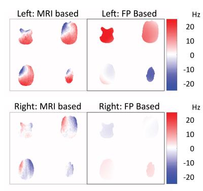
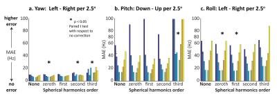
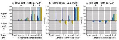
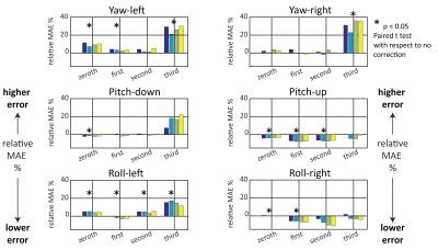
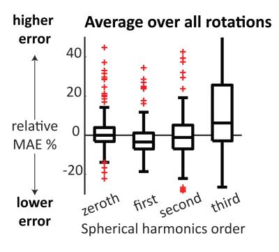
A comparison example is shown in Figure 2 between “ground-truth” measured B0 maps and FP based estimation of B0 for the case of yaw-to-the-left and yaw-to-the-right: difference B0 maps with respect to the ‘base’ position are also shown. Results from the B0 simulations for all three rotation axes are summarized in terms of the MAE, plotted in Figure 3. It was observed that a ‘yaw’ rotation in general has a lower absolute error in B0 (MAE 8 Hz) than Pitch (20 Hz) and Roll (30 Hz) regardless of the spherical harmonics (SH) order. The effect of the SH order per rotation axis is more evident in MAE relative to no correction after rotation (rMAE), depicted in Figure 4. The figure shows a significant increase in rMAE for yaw and pitch with third order SH in simulation. A similar significant increase for third order SH is seen for yaw rotation in in-vivo, shown in Figure 5. The rMAE and its dependence on spherical harmonic order is summarized in Figure 6, showing the smallest spread of rMAE for the first order SH and the median of its measurements has an rMAE lower than 0, indicating a decrease in error, while the third order has a large spread in rMAE with a median above 0.Discussion
When the head is rotated, correcting with FP estimated B0 maps can actually lead to an increase in the error in B0 estimation. This is most evident when using third order SH correction, although this shows a large spread in rMAEs albeit with a positive median (5% increase in error). However, when using the first order SH correction improved performance is identified since the median of rMAE rotation measurements becomes negative. When using zeroth or second order SH orders the median rMAE is approximately zero.Acknowledgements
No acknowledgement found.References
1. De Zanche N, Barmet C, Nordmeyer-Massner JA, Pruessmann KP. NMR probes for measuring magnetic fields and field dynamics in MR systems. Magn. Reson. Med. 2008;60:176–186.
2. Hess AT, Dylan Tisdall M, Andronesi OC, Meintjes EM, van der Kouwe AJW. Real-time motion and B0 corrected single voxel spectroscopy using volumetric navigators. Magn. Reson. Med. 2011;66:314–323. doi: 10.1002/mrm.22805.
3. Salomir R, de Senneville BD, Moonen CT. A fast calculation method for magnetic field inhomogeneity due to an arbitrary distribution of bulk susceptibility. Concepts Magn. Reson. Part B Magn. Reson. Eng. 2003;19B:26–34. doi: 10.1002/cmr.b.10083.
Figures
