3916
DREAM-based B1-shimming for cardiac imaging at 7 T1Division Imaging, Department of Radiology, University Medical Center Utrecht, Utrecht, Netherlands, 2Division Imaging, Department of Radiotherapy, University Medical Center Utrecht, Utrecht, Netherlands, 3Biomedical Image Analysis, Eindhoven University of Technology, Eindhoven, Netherlands
Synopsis
RF shimming for multi-transmit systems is commonly performed by phase-only shimming. Improved B1+ homogeneity is reached by phase-amplitude shimming but this requires knowledge of the B1+ magnitude distributions. For cardiac imaging at 7 Tesla, the acquisition of these distributions is challenging. We present a DREAM-based acquisition series to reconstruct B1+ magnitude maps in the heart. This method is applied to homogenize the transmit field for three subjects using phase-amplitude shimming. Results demonstrate a clear improvement of transmit field homogeneity in the heart in comparison to phase-only
Purpose
Cardiac imaging at 7 T holds promise for several clinical applications such as coronary imaging, functional imaging and quantitative T2-mapping1–4. One of the inherent challenges of ultrahigh field- (UHF-) MRI is the inhomogeneity of the transmit field5,6. Although commonly only phase shimming is applied, phase-amplitude shimming can improve the homogeneity of the transmit field in a region of interest such as the heart. The individual transmit profiles of all transmit channels should be known before phase-amplitude shimming can be applied. Several methods are available to measure or estimate these transmit profiles, but measuring transmit profiles in the heart is challenging7–9. In this work, a previously proposed method to measure transmit profiles based on a measured B1+-map is applied to cardiac imaging at ultrahigh-field8. The DREAM B1+-mapping method was used to measure B1+ within the time constraints of cardiac triggered imaging10. The new shimming method is applied to enhance the homogeneity of the transmit field in the heart, and it is compared to phase-only shimming.Methods
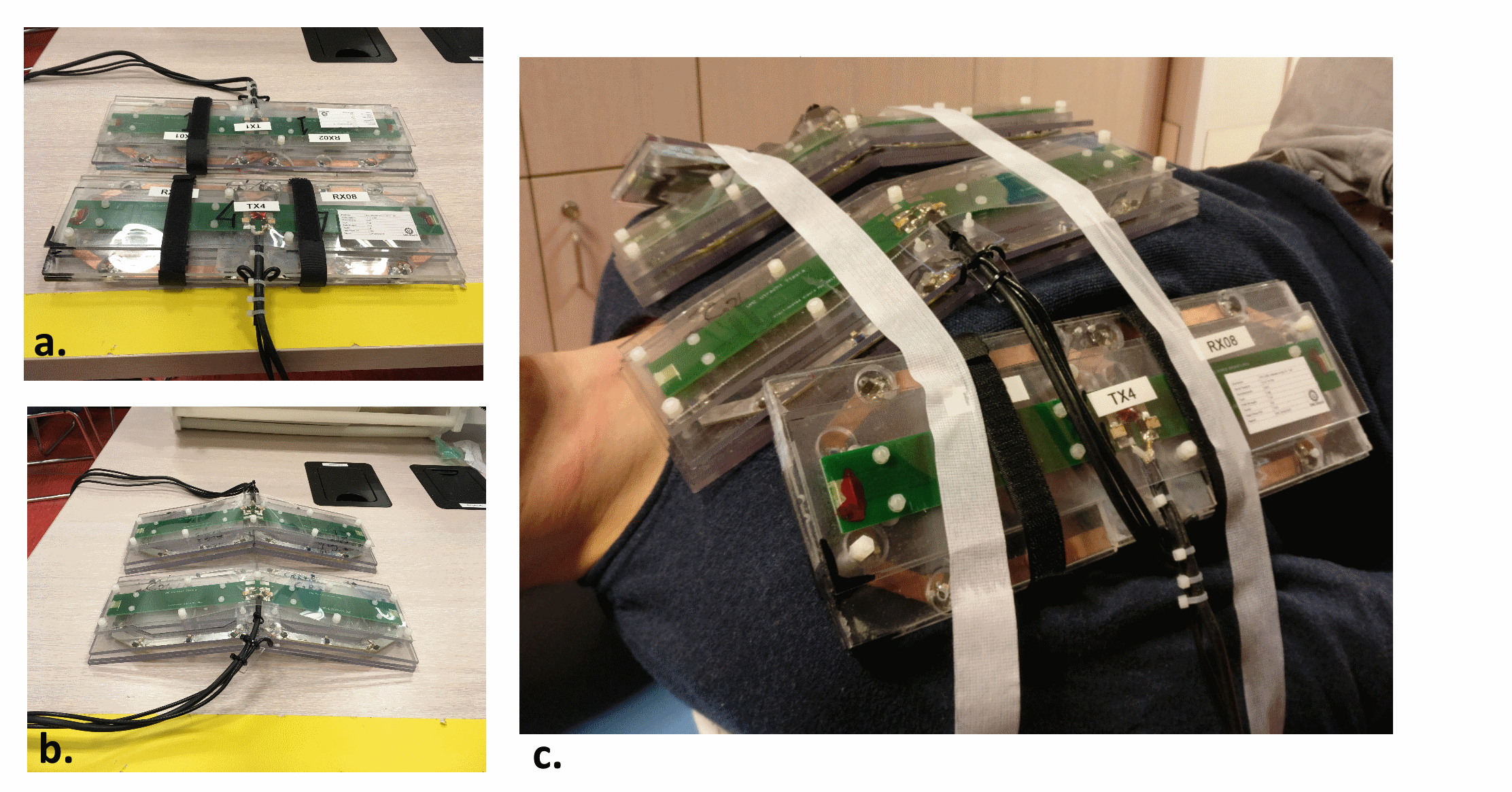
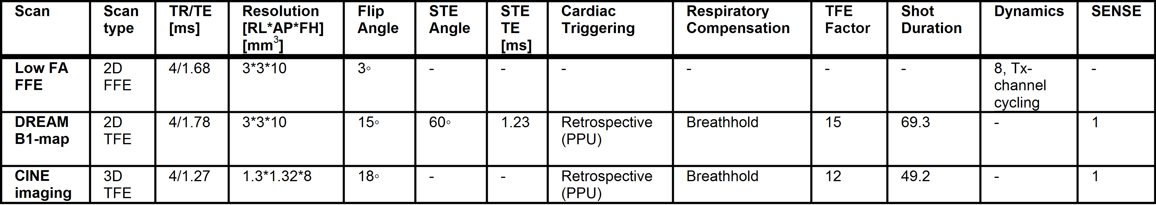
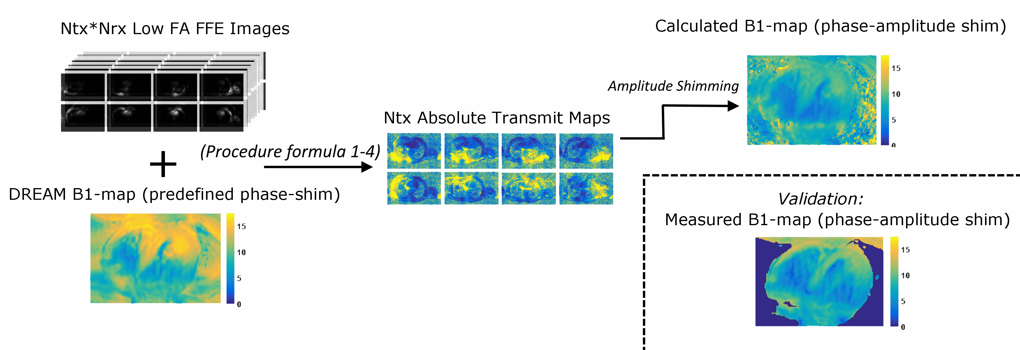
A 8 channel Tx-/24 channel Rx-setup (figure 1) was used for cardiac imaging at 7 T (Philips Healthcare, Best)11 on three healthy volunteers. Informed consent was obtained following the protocols of our institutional review board. The scan parameters of all sequences used for this abstract are shown in figure 2. A series of low flip angle FFE-images (ρB1,i+B1,j-) was obtained for all Ntx transmit channels and reconstructed separately for all Nrx receive channels. Also, a DREAM B1+-map (B1+shimmed,DREAM) was obtained with predefined phase-shimmed settings wi. Nrx low flip angle FFE-images with shim settings wi were reconstructed from the low flip angle FFE images [1]. The spin density/receive profile of channel j is reconstructed by dividing the nRx images of [1] by B1+shimmed,DREAM [2]. Now for every receive channel j, the transmit profiles for every transmit channel i can be reconstructed [3]. Final transmit profiles are calculated as a weighted sum of the transmit profiles for each receive channel j [4].
[1] $$\rho B_{1,shimmed}^+B_{1,j}^-=\sum_1^{i=Ntx}w_i\rho B_{1,i}^+ B_{1,j}^-$$
[2] $$\rho B_{1,j}^- = \frac{\rho B_{1,shimmed}^+B_{1,j}^-}{B_{1,shimmed,DREAM}^+}$$
[3] $$B_{1,i,j}^+=\frac{\rho B_{1,i}^+B_{1,j}^-}{\rho B_{1,j}^-}$$
[4] $$B_{1,i}^+=\sum_1^{j=Nrx}\frac{\rho B_{1,shimmed}^+B_{1,j}^-(B_{1,i,j}^+)}{\rho B_{1,shimmed}^+B_{1,j}^-}$$
Based on the absolute transmit profiles, phase amplitude-shimming was applied to minimize the signal coefficient of variation in the heart. A graphic chart of this procedure is shown in figure 3. B1+-maps were obtained after phase-amplitude shimming and after phase-only shimming where only the phase distributions were included as an input (no amplitude information). The B1+-maps are compared in terms of coefficient of variation (CoV) and magnitude. Additionally, functional CINE images were obtained with both shim settings and compared based on homogeneity.
Results
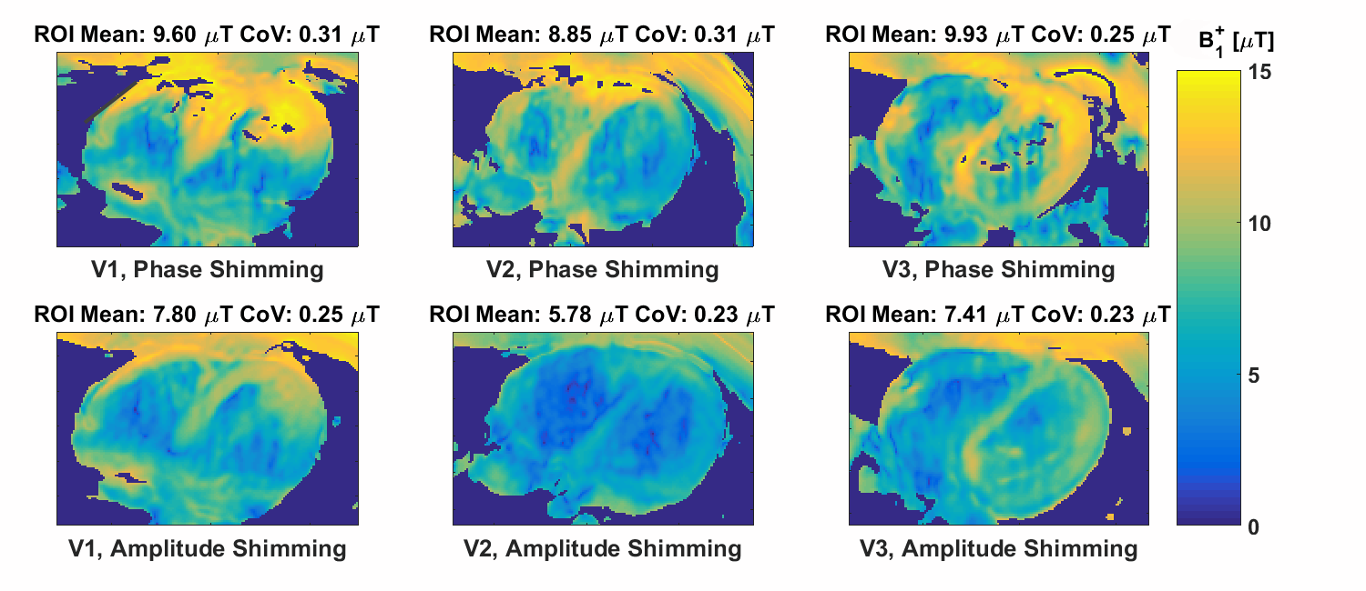
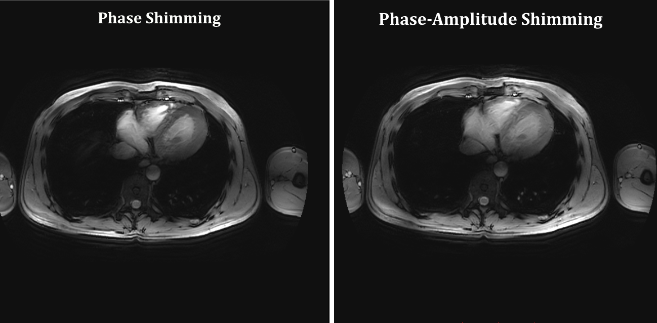
Figure 4 shows the B1+-maps that were obtained for both shim methods. In a total of 3 volunteers, the average CoV was decreased from 0.29 to 0.236, which is a decrease of 23%. The average B1+ in the cardiac region is decreased from 9.4 to 7 μT. However, due to amplitude scaling, the input power is also lowered from 6.4 kW to 3.2 kW. The B1+-efficiency is 0.12 μT/√W in both cases. Figure 5 shows a slice of a CINE image obtained on the same volunteer for both phase-only shimming and phase-amplitude shimming. In the phase shimmed image, the signal intensity is very high at the anterior side of the heart, while for the phase-amplitude shimming approach the signal is more homogeneous throughout the heart.Discussion
It is demonstrated that by utilizing a DREAM-based reconstruction of transmit profiles and phase-amplitude shimming, the CoV of B1+ in the heart can be decreased by an average of 23%, while transmit efficiency is not affected negatively. By decreasing the total peak power, local SAR may be lowered, this still needs to be investigated. The DREAM B1+-maps that are shown in figure 2 may be further improved by additional measures such as flow compensation and fitting of the B1+-signal which could improve results even more12,13.Conclusion
A DREAM based method is presented to reconstruct the single channel B1+ magnitude distributions for cardiac imaging at 7 Tesla. It is demonstrated that by using this information for phase-amplitude shimming, the coefficient of variation in the heart can be decreased by an average of 23% in 3 volunteers, leading to a visible improvement in image quality.Acknowledgements
This work is part of a project funded by Dutch Technology Foundation STW, project number 13783References
1. Bizino MB, Bonetti C, Geest RJ Van Der, Versluis MJ, Webb AG, Lamb HJ. High Spatial Resolution Coronary Magnetic Resonance Angiography at 7 T Comparison With Low Spatial Resolution Bright Blood Imaging. 2014;49(5).
2. Niendorf T, Paul K, Oezerdem C, Graessl A, Klix S, Huelnhagen T, Hezel F, Rieger J, Waiczies H, Frahm J, et al. W ( h ) ither human cardiac and body magnetic resonance at ultrahigh fields?? technical advances , practical considerations , applications , and clinical opportunities. 2015;(October 2014).
3. Raaijmakers AJE, Aidi H El, Versluis M, Webb A, Lamb HJ, Luijten PR, Berg CAT Van Den, Leiner T. Proc. Intl. Soc. Mag. Reson. Med. 22 (2014) 4923. 2014;7:4923.
4. Volunteers YH, Versluis MJ, Westenberg JJM, Smith NB, Stuber M, Roos A De, Webb AG. Right Coronary MR Angiography at 7 T: A Direct Quantitative and Qualitative Comparison with 3 T, Radiology 2010;257(1).
5. Vaughan JT, Garwood M, Collins CM, Liu W, DelaBarre L, Adriany G, Andersen P, Merkle H, Goebel R, Smith MB, et al. 7T vs. 4T: RF power, homogeneity, and signal-to-noise comparison in head images. Magnetic resonance in medicine. 2001;46(1):24–30.
6. Metzger GJ, Snyder C, Akgun C, Vaughan T, Ugurbil K, de Moortele V, others. Local B1+ shimming for prostate imaging with transceiver arrays at 7T based on subject-dependent transmit phase measurements. Magnetic Resonance in Medicine. 2008;59(2):396–409.
7. Van de Moortele PF UK. Very Fast Multi Channel B1 Calibration at High Field in the Small Flip Angle Regime. Proceedings of the 17th Annual Meeting of ISMRM, Honolulu, USA. 2009;3:367.
8. Van De Moortele P-F;Snyder C. Calibration Tools for RF Shim at Very High Field with Multiple Element RF Coils: From Ultra Fast Local Relative Phase to Absolute Magnitude B1+ Mapping. Ismrm. 2007;15:1.
9. Padormo F, Hess AT, Aljabar P, Malik SJ, Jezzard P, Robson MD, Hajnal J V., Koopmans PJ. Large dynamic range relative B1+ mapping. Magnetic Resonance in Medicine. 2016;76(2):490–499.
10. Nehrke K, Boernert P. DREAM-a novel approach for robust, ultrafast, multislice B1 mapping. Magnetic Resonance in Medicine. 2012;68(5):1517–1526.
11. Voogt IJ, Klomp DWJ, Hoogduin H, Luttje MP, Luijten PR, van den Berg CAT, Raaijmakers AJE. Combined 8-channel transceiver fractionated dipole antenna array with a 16-channel loop coil receive array for body imaging at 7 Tesla. Ismrm 2015. 2015;200(2007):631.
12. Nehrke K, Sprinkart AM, Schild HH, Börnert P. Fast B1+ Mapping for Cardiac MR using a Black Blood DREAM Sequence. Ismrm. 2013;21(c):4271.
13. Sbrizzi A, Hoogduin H, Lagendijk JJ, Luijten P, Van Den Berg CAT. Robust reconstruction of B1+ maps by projection into a spherical functions space. Magnetic Resonance in Medicine. 2014;71(1):394–401.
Figures